. 2012 May 16;31(10):2261-74.
doi: 10.1038/emboj.2012.79. Epub 2012 Apr 13.

The mechanism of γ-Secretase dysfunction in familial Alzheimer disease

Affiliations

The mechanism of γ-Secretase dysfunction in familial Alzheimer disease

Lucía Chávez-Gutiérrez et al. EMBO J. .

Abstract

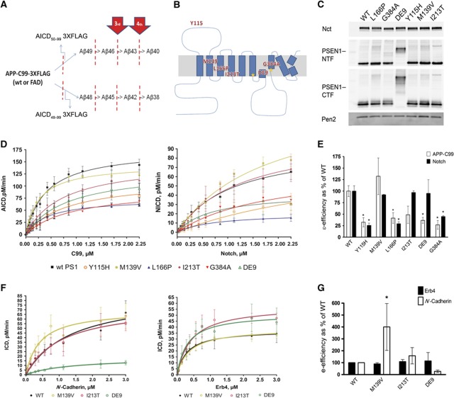
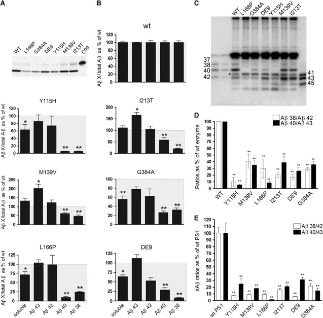
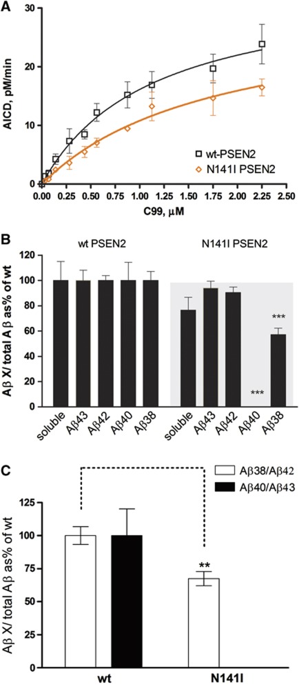
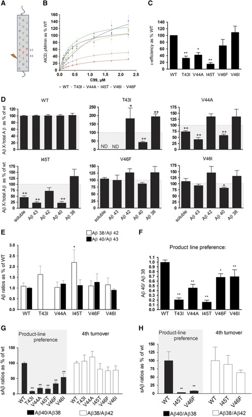
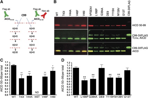
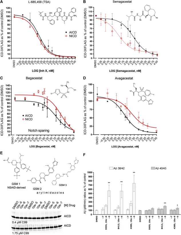
The mechanisms by which mutations in the presenilins (PSEN) or the amyloid precursor protein (APP) genes cause familial Alzheimer disease (FAD) are controversial. FAD mutations increase the release of amyloid β (Aβ)42 relative to Aβ40 by an unknown, possibly gain-of-toxic-function, mechanism. However, many PSEN mutations paradoxically impair γ-secretase and 'loss-of-function' mechanisms have also been postulated. Here, we use kinetic studies to demonstrate that FAD mutations affect Aβ generation via three different mechanisms, resulting in qualitative changes in the Aβ profiles, which are not limited to Aβ42. Loss of ɛ-cleavage function is not generally observed among FAD mutants. On the other hand, γ-secretase inhibitors used in the clinic appear to block the initial ɛ-cleavage step, but unexpectedly affect more selectively Notch than APP processing, while modulators act as activators of the carboxypeptidase-like (γ) activity. Overall, we provide a coherent explanation for the effect of different FAD mutations, demonstrating the importance of qualitative rather than quantitative changes in the Aβ products, and suggest fundamental improvements for current drug development efforts.


Conflict of interest statement

Harrie Gijsen is an employee of Janssen Pharmaceutica. Bart de Strooper is a consultant for Janssen Pharmaceutica, Envivo Pharmaceutics and Remynd and is supported by research grants from Janssen Pharmaceutics.

Figures

Figure 1
Figure 1
FAD–PSEN1 mutations do not consistently decrease the enzymatic efficiency of the endopeptidase cleavage. (A, B) Schematic overviews of APP processing and location of FAD–PSEN1 mutations used in the current study. (C) Expression levels of Nct, PSEN1–NTF, PSEN1–CTF and Pen-2 in Psen1/2−/− mEFs transduced with human wt or FAD–PSEN1 mutants using a replication-defective recombinant retroviral expression system (Clontech) and selected with puromycin (5 μg/ml). Western blotting and densitometric analysis of the CHAPSO-solubilized membrane proteins from the different PSEN1 cell lines indicate that wt and mutant PSEN1 rescued γ-secretase complex to similar extents. In order to determine specific activities for the wt or FAD complexes, γ-secretase activities were normalized to PSEN CTF fragment levels or full-length PS1 levels for the DE9 mutant. (D) Kinetic curves for wt and PS1–FAD mutants using purified APP-C99-3XFLAG or Notch-3XFLAG substrates (mean±s.e.) or (F) ErB4-3XFLAG and N-Cadherin-3XFLAG substrates (mean±s.d.). Detergent-extracted membranes were incubated in 0.25% CHAPSO reaction buffer with varying concentrations of purified substrate for 4 h at 37 °C. In vitro generated ICD-3XFLAG were analysed by quantitative western blot analysis (see Materials and methods). (E) FAD–PSEN1 ε-enzymatic efficiencies for APP-C99 and Notch substrates (mean±s.e.). Enzymatic efficiencies unequivocally demonstrate that loss of function at the ε-cleavage is not a constant among PSEN1 mutations. (G) FAD–PSEN1 mutations that did not affect the generation of NICD did not change significantly the processing of ErB4 (mean±s.d.) either. In contrast, N-Cadherin processing was significantly upregulated by the M139V (mean±s.d.). (E, G) Experiments were repeated 3–5 times. Statistical significance of the data was tested with one-way analysis of variance (ANOVA) and Dunnett’s post test, taking the corresponding WT efficiency as control group, *P<0.05.
Figure 2
Figure 2
FAD–PSEN1 mutations impair the fourth enzymatic turnover. AICD levels (moles per min) generated by the wt or FAD mutant complexes (A) were used to normalize Aβ products (moles per min) in order to determine accurately Aβ generation relative to C99 substrate. Aβ profiles (B) thus represent Aβ products corrected for the initial endoprotease activities, plotted as percentage of the wt Aβ products (mean±s.e.). Soluble Aβ (sum of Aβ38, Aβ40, Aβ42 and Aβ43 peptides) gives information about the efficiency of the γ-cleavages: lower levels (<100%, grey box) suggest that longer peptides (>Aβ43) accumulate in the reactions. (C) In agreement with the ELISA quantifications, total Aβ analysed in urea-based gels show increments in Aβ42 and Aβ43, and reductions in Aβ40 and Aβ38 in FAD–PSEN1 mutations, relative to wt. (*) Indicates a non product band that is present in the C99 substrate (see Supplementary Figure 2). (D) Aβ product/substrate ratios determined in vitro for the FAD–PSEN1 mutations show an impairment at the fourth γ-secretase turnover (mean±s.e.). Experiments in (B) and (D) were repeated 4–6 times. Statistical significance of the data tested with one-way ANOVA and Dunnett’s post test taking the corresponding WT as control group; *P<0.05, **P<0.01. (E) Aβ product/substrate ratios determined in vivo confirm impairment at the fourth enzymatic cycle: wt or FAD–PSEN1 mEF cell lines were transiently transduced with APPswe, extracellular media collected at 24 h after infection and sAβ measured by ELISA (mean±s.e.). Statistical significance: n=4, ANOVA and Dunnett’s post test, **P<0.01.
Figure 3
Figure 3
FAD–PSEN2 N141I impair the fourth enzymatic turnover. (A) Kinetic curves for wt and PSEN2–FAD N141I mutant using purified APP-C99-3XFLAG as substrate (mean±s.e.). (B) Aβ profiles represent Aβ products corrected for the initial endoprotease activities, plotted as % of the wt Aβ products (mean±s.e.). Soluble Aβ (sum of Aβ38, Aβ40, Aβ42 and Aβ43 peptides) suggests accumulation of longer peptides (>Aβ43) in the mutant reactions. (C) Aβ product/substrate ratios determined in vitro for the FAD–PSEN2 mutation show an impairment at the fourth γ-secretase turnover (mean±s.e.). In (B) and (C) statistical significance (two-tailed t-test) is indicated by **P<0.005 and ***P<0.001. Note that N141I abolishes Aβ40 generation.
Figure 4
Figure 4
FAD substrate mutations shift APP processing towards the Aβ38 product line. (A) Schematic overview of FAD–APP mutations used in this study. (B) Kinetic curves for the ε-processing of APP. Detergent-extracted membranes from Psen1/2−/− mEFs rescued with human wt PSEN1 were incubated in 0.25% CHAPSO reaction buffer, with varying concentrations of purified wt or FAD–APP substrates. AICD-3XFLAG levels were analysed by quantitative western blot analysis (see Materials and methods). (C) Enzymatic efficiencies for FAD–APP-C99 substrates (mean±s.e.) prove that AICD generation is affected in three out of five FAD-mutant substrates. (D) FAD Aβ product profiles suggest consistent increments in Aβ42 and Aβ38. Soluble Aβ levels (sum of Aβ38, Aβ40, Aβ42 and Aβ43 peptides) suggest accumulations of longer Aβ peptides in the γ-processing of the V44A and I45T mutants. The T43I mutation disrupts the epitope for the anti-Aβ43-specific antibody, thus neither Aβ43 nor soluble Aβ levels could be determined (ND). (E) Aβ product/substrate ratios reveal that APP mutations do not consistently affect the fourth γ-secretase turnover, but change the product-line preference as indicated by the Aβ40/Aβ38 ratio (F). (G) sAβ in the conditioned media of HEK293 cells transiently transfected with wt or FAD-C99 mutants were quantified by ELISA (see Supplementary Figure 4). sAβ ratios indicate that APP–FAD substrate mutants change the product-line preference towards the Aβ48>Aβ38 (Aβ40/Aβ38) in living cells, but do not affect the fourth catalytic turnover of the γ-secretase (Aβ38/Aβ42) (mean±s.e., n=5). (H) Primary cultured neurons were transduced with SFV expressing WT APP or the indicated mutant substrates (mean±s.d., n=3). (CH) Statistical significance tested with one-way ANOVA and Dunnett’s post-test, taking the corresponding WT as control group; *P<0.05,**P<0.01.
Figure 5
Figure 5
Shift in the ε-cleavage position contributes to the FAD-associated phenotype. (A) Detection of AICD50–99 and total AICD using a neo-epitope antibody and the FLAG-M2 antibody, respectively. (B) SDS–PAGE/western blot analysis of AICD products from either wt and FAD substrates (left panel) or wt and FAD–PSEN1mutants (right panel). Signals for the AICD50–99 neo-epitope antibody or the FLAG-M2 antibody are shown in red and green, respectively. Overlapping neo-epitope and FLAG antibody signals are displayed in yellow. (C) AICD50–99/total AICD ratios indicate that FAD–APP mutations promote the Aβ38 product line by shifting the ε-cleavage position. The I45T could not be included in the analysis because of extremely low AICD signals (ND, not determined). (D) This pathogenic mechanism is also observed in some FAD–PSEN1 mutations. Statistical significance of the data (n=5) tested with ANOVA and Dunnett’s post test, taking AICD generated in WT conditions as control group; *P<0.05,**P<0.01.
Figure 6
Figure 6
Analysis of GSI and GSM. Dose-response inhibitory assays for (A) the transition state analogue (TSA) L-685,458 (InhX), (B) semagacestat, and the Notch-sparing compounds (C) begacestat and (D) avagacestat (see materials in Supplementary data) were performed using CHAPSO-extracted membranes from dKO PSEN1/2 MEFs stably expressing human wt PSEN1 as source of γ-secretase and 1 × Km substrate concentrations (400 nM APP-C99-3XFLAG or 1 μM Notch-3XFLAG). Structures of the different compounds are displayed. In vitro-generated AICD (in black) or NICD (in red) are plotted as percentage of control reaction (DMSO). Error bars indicate s.d. (n=3); except for semagacestat plot (s.e., n=5). (E) Top panel: structures of the GSM tested. Low panel: increasing concentrations of GSM 1–3 did not change in vitro AICD generation, neither at 0.4 μM APP-C99 substrate (1 × Km) nor at saturating conditions (1.75 μM C99-3XFLAG). (F) Effect of increasing concentrations of GSM 1–3 on Aβ production at 1 × Km APP-C99 substrate (0.4 μM): Aβ product/substrate ratios show that GSM 1–3 specifically activate the fourth cycle of the γ-secretase complex. In particular, GSM activate the Aβ38 product line. Panel shows mean±s.e.; statistical significance of the data (n=4) tested with ANOVA and Dunnett’s post test, vehicle (DMSO) as control group; *P<0.05,**P<0.01.

Comment in

References

    1. Annaert WG, Esselens C, Baert V, Boeve C, Snellings G, Cupers P, Craessaerts K, De Strooper B (2001) Interaction with telencephalin and the amyloid precursor protein predicts a ring structure for presenilins. Neuron 32: 579–589 - PubMed
    1. Annaert WG, Levesque L, Craessaerts K, Dierinck I, Snellings G, Westaway D, George-Hyslop PS, Cordell B, Fraser P, De Strooper B (1999) Presenilin 1 controls gamma-secretase processing of amyloid precursor protein in pre-golgi compartments of hippocampal neurons. J Cell Biol 147: 277–294 - PMC - PubMed
    1. Bentahir M, Nyabi O, Verhamme J, Tolia A, Horre K, Wiltfang J, Esselmann H, De Strooper B (2006) Presenilin clinical mutations can affect gamma-secretase activity by different mechanisms. J Neurochem 96: 732–742 - PubMed
    1. Berezovska O, Lleo A, Herl LD, Frosch MP, Stern EA, Bacskai BJ, Hyman BT (2005) Familial Alzheimer’s disease presenilin 1 mutations cause alterations in the conformation of presenilin and interactions with amyloid precursor protein. J Neurosci 25: 3009–3017 - PMC - PubMed
    1. Bergmans BA, De Strooper B (2010) Gamma-secretases: from cell biology to therapeutic strategies. Lancet Neurol 9: 2215–226 - PubMed

Publication types

MeSH terms