. 2012 Apr;68(Pt 4):431-40.
doi: 10.1107/S090744491200251X. Epub 2012 Mar 17.

JLigand: a graphical tool for the CCP4 template-restraint library

Affiliations

JLigand: a graphical tool for the CCP4 template-restraint library

Andrey A Lebedev et al. Acta Crystallogr D Biol Crystallogr. 2012 Apr.

Abstract

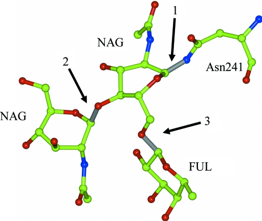
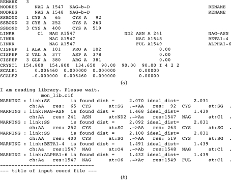
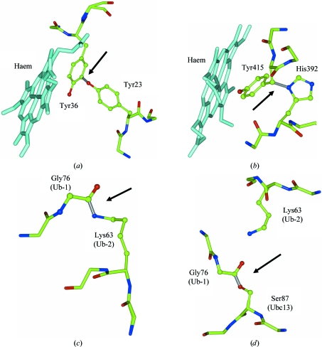
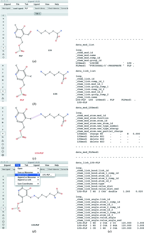
Biological macromolecules are polymers and therefore the restraints for macromolecular refinement can be subdivided into two sets: restraints that are applied to atoms that all belong to the same monomer and restraints that are associated with the covalent bonds between monomers. The CCP4 template-restraint library contains three types of data entries defining template restraints: descriptions of monomers and their modifications, both used for intramonomer restraints, and descriptions of links for intermonomer restraints. The library provides generic descriptions of modifications and links for protein, DNA and RNA chains, and for some post-translational modifications including glycosylation. Structure-specific template restraints can be defined in a user's additional restraint library. Here, JLigand, a new CCP4 graphical interface to LibCheck and REFMAC that has been developed to manage the user's library and generate new monomer entries is described, as well as new entries for links and associated modifications.


Figures

Figure 1
Figure 1
Example of glycosylation, which only requires the generic links for sugars defined in REFMAC’s standard monomer library for refinement. The figure shows one molecule of β-l-fucose (FUL) and two molecules of N-­acetyl-d-glucosamine (NAG) forming a polysaccharide covalently bound to Asn241 in the crystal structure of the G117H mutant of human butyrylcholinesterase (Nachon et al., 2011; PDB entry 2xmb). Refinement of such a structure will implicitly use the links NAG-ASN (1), BETA1-4 (2), ALPHA1-6 (3) and associated modification from the restraint library. This figure and Fig. 3 ▶ were generated using CCP4mg (Potterton et al., 2004 ▶).
Figure 2
Figure 2
REFMAC checkpoints for refinement of glycosylated proteins. The figure shows fragments of (a) the output PDB file and (b) the REFMAC log file after successful refinement of the structure presented in Fig. 1 ▶. LINKR records indicate that REFMAC was able to associate short distances between monomers with library link entries and corresponding warnings in the log file prompt the user to check whether this assignment is correct and whether the linked atoms fit the electron density.
Figure 3
Figure 3
Examples of side chain to side chain and side chain to main chain covalent linkages in proteins (see citations and PDB codes in the main text): (a) Tyr–Tyr covalent link in M. tuberculosis haemoglobin O. (b) Tyr–His covalent link in HPII from E. coli. In both (a) and (b) the new covalent bond increases the rigidity of the haem site. (c) Lys to main chain (C-terminal Gly) link between ubiquitin molecules in the complex of the TAB2 protein with diubiquitin. TAB2 only binds linked ubiquitin molecules; the interaction is not shown, being distant from the link. (d) Covalent link between ubiquitin main chain and the side chain of Ser from the ubiquitin-conjugation enzyme Ubc13. Lys63 of the second ubiquitin is poised to attack the intermediate ester bond. Residues involved in covalent-linkage formation are shown in ball-and-stick representation. The linkages are shown in grey and are indicated by arrows.
Figure 4
Figure 4
Defining restraints for the covalent linkage Lys–PLP using JLigand. Snapshots show the state of the JLigand interface after the following user actions: (a) loading monomers LYS and PLP from the restraint library, (b) removing O4A from PLP, (c) connecting C4A (PLP) with NZ (LYS) and defining the bond order and (d) regularization. The Save As Link menu has to be used to generate (e) an additional library file containing the following five data blocks: list of modifications, list of links, modifications LYSmod1 and PLPmod1 to be applied to LYS and PLP, respectively, and link LYS–PLP. The file header, tables of restraints for LYSmod1 and all the tables for PLPmod1 are omitted, as indicated by lines filled by tildes. H atoms are dealt with automatically. It is also possible to visualize them and handle them explicitly.

Similar articles

Cited by

References

    1. Bernstein, F. C., Koetzle, T. F., Williams, G. J., Meyer, E. F. Jr, Brice, M. D., Rodgers, J. R., Kennard, O., Shimanouchi, T. & Tasumi, M. (1977). J. Mol. Biol. 112, 535–542. - PubMed
    1. Brady, J. D., Sadler, I. H. & Fry, S. C. (1996). Biochem. J. 315, 323–327. - PMC - PubMed
    1. Bravo, J., Fita, I., Ferrer, J. C., Ens, W., Hillar, A., Switala, J. & Loewen, P. C. (1997). Protein Sci. 6, 1016–1023. - PMC - PubMed
    1. Bricogne, G., Blanc, E., Brandl, M., Flensburg, C., Keller, P., Paciorek, W., Roversi, P., Sharff, A., Smart, O. S., Vonrhein, C. & Womack, T. O. (2011). BUSTER Cambridge: Global Phasing Ltd.
    1. Collaborative Computational Project, Number 4 (1994). Acta Cryst. D50, 760–763. - PubMed

Publication types