

Extratemporal functional connectivity impairments at rest are related to memory performance in mesial temporal epilepsy
- PMID: 22505284
- PMCID: PMC3864618
- DOI: 10.1002/hbm.22059
Extratemporal functional connectivity impairments at rest are related to memory performance in mesial temporal epilepsy
Abstract
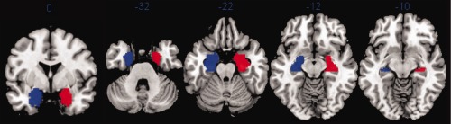
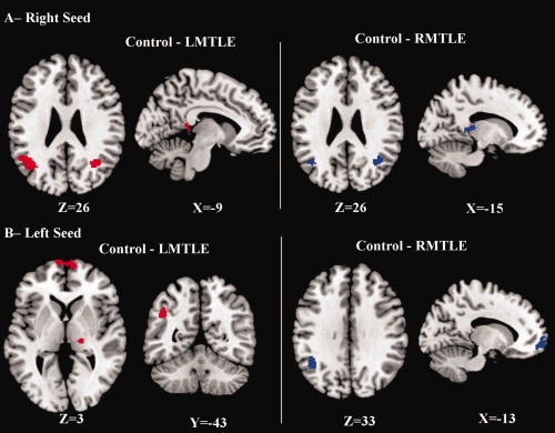
Mesial temporal lobe epilepsy (MTLE) is the most frequent form of focal epilepsy. At rest, there is evidence that brain abnormalities in MTLE are not limited to the epileptogenic region, but extend throughout the whole brain. It is also well established that MTLE patients suffer from episodic memory deficits. Thus, we investigated the relation between the functional connectivity seen at rest in fMRI and episodic memory impairments in MTLE. We focused on resting state BOLD activity and evaluated whether functional connectivity (FC) differences emerge from MTL seeds in left and right MTLE groups, compared with healthy controls. Results revealed significant FC reductions in both patient groups, localized in angular gyri, thalami, posterior cingulum and medial frontal cortex. We found that the FC between the left non-pathologic MTL and the medial frontal cortex was positively correlated with the delayed recall score of a non-verbal memory test in right MTLE patients, suggesting potential adaptive changes to preserve this memory function. In contrast, we observed a negative correlation between a verbal memory test and the FC between the left pathologic MTL and posterior cingulum in left MTLE patients, suggesting potential functional maladaptative changes in the pathologic hemisphere. Overall, the present study provides some indication that left MTLE may be more impairing than right MTLE patients to normative functional connectivity. Our data also indicates that the pattern of extra-temporal FC may vary as a function of episodic memory material and each hemisphere's capacity for cognitive reorganization.
Keywords: connectivity; epilepsy; episodic memory; fMRI; functional reorganization; resting state.
Copyright © 2012 Wiley Periodicals, Inc., a Wiley company.
Figures


References
-
- Alessio A, Damasceno BP, Camargo CH, Kobayashi E, Guerreiro CA, Cendes F (2004): Differences in memory performance and other clinical characteristics in patients with mesial temporal lobe epilepsy with and without hippocampal atrophy. Epilepsy Behav 5:22–27. - PubMed
-
- Alessio A, Pereira FR, Sercheli MS, Rondina JM, Ozelo HB, Bilevicius E, Pedro T, Covolan RJ, Damasceno BP, Cendes F (2011): Brain plasticity for verbal and visual memories in patients with mesial temporal lobe epilepsy and hippocampal sclerosis: An fMRI study. Hum Brain Mapp. doi: 10.1002/hbm.21432. - PMC - PubMed
-
- Arnold S, Schlaug G, Niemann H, Ebner A, Luders H, Witte OW, Seitz RJ (1996): Topography of interictal glucose hypometabolism in unilateral mesiotemporal epilepsy. Neurology 46:1422–1430. - PubMed
-
- Bettus G, Bartolomei F, Confort‐Gouny S, Guedj E, Chauvel P, Cozzone PJ, Ranjeva JP, Guye M (2010): Role of resting state functional connectivity MRI in presurgical investigation of mesial temporal lobe epilepsy. J Neurol Neurosurg Psychiatry 81:1147–1154. - PubMed
Publication types
MeSH terms
Grants and funding
LinkOut - more resources
Full Text Sources
Medical
