. 2012 May;122(5):1628-43.
doi: 10.1172/JCI60660. Epub 2012 Apr 16.

The DC receptor DNGR-1 mediates cross-priming of CTLs during vaccinia virus infection in mice

Affiliations

The DC receptor DNGR-1 mediates cross-priming of CTLs during vaccinia virus infection in mice

Salvador Iborra et al. J Clin Invest. 2012 May.

Abstract

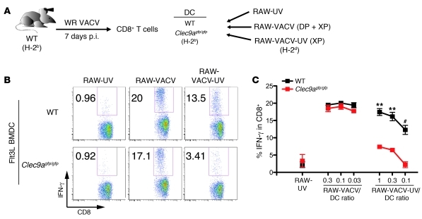
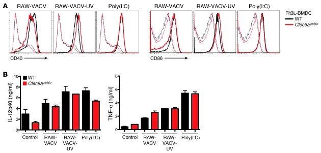
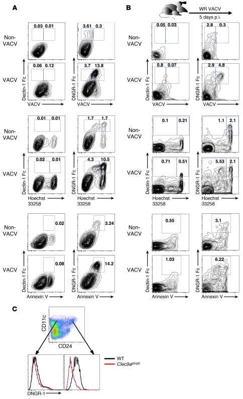
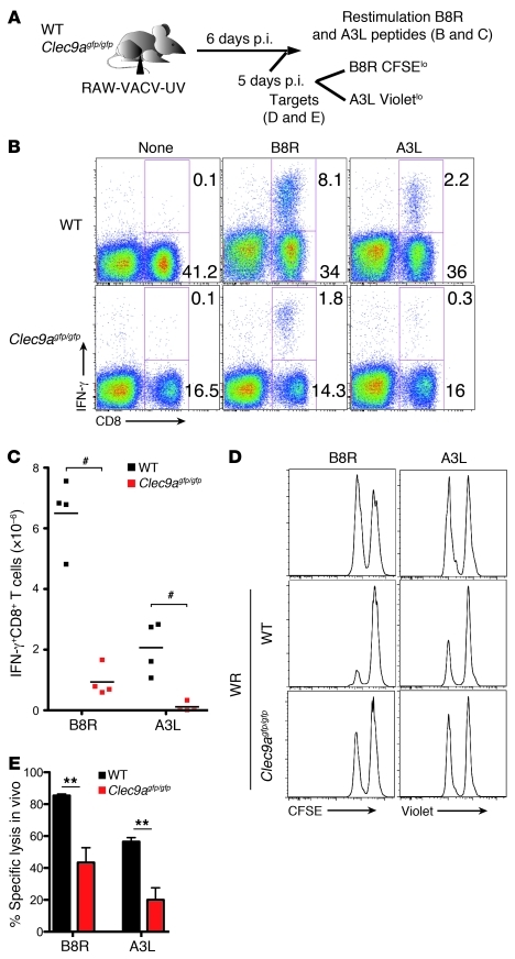
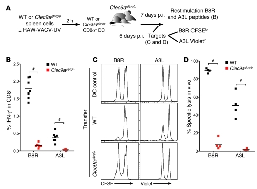
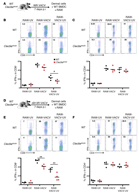
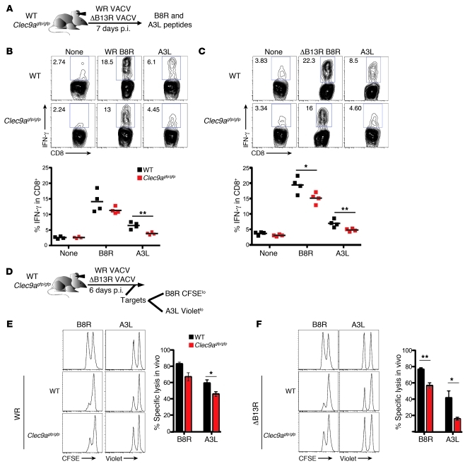
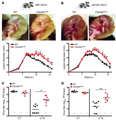
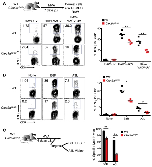
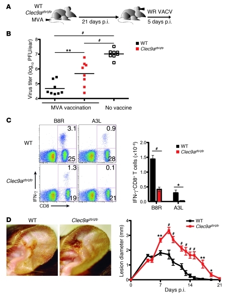
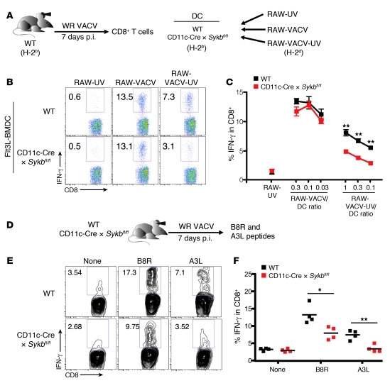
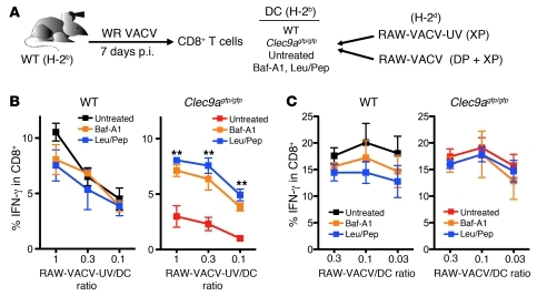
In order to prime T cells, DCs integrate signals emanating directly from pathogens and from their noxious action on the host. DNGR-1 (CLEC9A) is a DC-restricted receptor that detects dead cells. Therefore, we investigated the possibility that DNGR-1 affects immunity to cytopathic viruses. DNGR-1 was essential for cross-presentation of dying vaccinia virus-infected (VACV-infected) cells to CD8(+) T cells in vitro. Following injection of VACV or VACV-infected cells into mice, DNGR-1 detected the ligand in dying infected cells and mediated cross-priming of anti-VACV CD8(+) T cells. Loss of DNGR-1 impaired the CD8+ cytotoxic response to VACV, especially against those virus strains that are most dependent on cross-presentation. The decrease in total anti-VACV CTL activity was associated with a profound increase in viral load and delayed resolution of the primary lesion. In addition, lack of DNGR-1 markedly diminished protection from infection induced by vaccination with the modified vaccinia Ankara (MVA) strain. DNGR-1 thus contributes to anti-VACV immunity, following both primary infection and vaccination. The non-redundant ability of DNGR-1 to regulate cross-presentation of viral antigens suggests that this form of regulation of antiviral immunity could be exploited for vaccination.


Figures

Figure 1
Figure 1. Lack of DNGR-1 blocks cross-presentation of vaccinia antigens in infected cells.
(AC) Production of IFN-γ by VACV-specific CD8+ effector T cells in response to cross-presented vaccinia antigens is severely impaired by the absence of DNGR-1 in Flt3L BMDCs. (A) For antigen presentation, RAW cells were either UV irradiated without infection (RAW-UV), infected with VACV without UV irradiation (RAW-VACV) to allow both direct presentation (DP) and cross-presentation (XP), or infected and subsequently irradiated to inactivate the virus (RAW-VACV-UV) to allow only cross-presentation. After 16 hours, RAW cells were exposed to Flt3L BMDCs from WT or Clec9agfp/gfp mice. As a readout of the restimulation ability of DCs, IFN-γ production was measured in CD8+ T cells from lymphoid organs of WT mice i.d. injected with WR VACV. (B) Representative set of dot plots. (C) Production of IFN-γ (mean ± SEM) from a representative experiment (n = 3 biological replicates) of 3 performed. **P < 0.01, #P < 0.001, unpaired 2-tailed Student’s t test.
Figure 2
Figure 2. DNGR-1 does not affect DC activation in response to vaccinia-infected cells.
Flt3L BMDCs from WT or Clec9agfp/gfp mice were untreated (control) or exposed for 20 hours to RAW-VACV or RAW-VACV-UV. (A) Induction of co-stimulatory molecules in DCs upon exposure to VACV-infected cells is not affected by the absence of DNGR-1. A representative set of histogram overlays is shown for CD40 (left) and CD86 (right). (B) Cytokine production induced in DCs by vaccinia-infected cells is not affected by the absence of DNGR-1. Data shown are from a representative experiment of 3 performed and are presented as mean ± SEM (n = 3 biological replicates). Differences were not statistically significant.
Figure 3
Figure 3. Cells infected with vaccinia virus expose DNGR-1 ligand in vitro and in vivo.
(A) Infection with VACV in vitro exposes DNGR-1 ligand. EL-4 cells were infected with WR VACV for 48 hours and stained with anti-vaccinia antibody and with control (Dectin-1–Fc) or DNGR-1–Fc constructs to detect the ligand and counterstained with annexin V and Hoechst 33258 (5 μg/ml). (B) Infection of C57BL/6 mice with VACV exposes DNGR-1 ligand simultaneously with vaccinia antigens in infected ear cells. WR VACV was injected i.d. in the ear, and, after 5 days, dermal cell suspensions were prepared as indicated in Methods. Staining and data are displayed as in A. (C) DNGR-1 is expressed in DCs locally in the ear. Cell suspensions from the ears of WT or Clec9agfp/gfp mice were semi-purified for CD11c+ cells by positive selection and stained for CD11c, CD24, and DNGR-1. A subset of CD11c+CD24hi DCs expresses DNGR-1. The dot plots in all panels are a replicate set of 3 from a representative experiment of 3 performed.
Figure 4
Figure 4. Lack of DNGR-1 blocks cross-presentation of VACV antigens in vivo.
(A) RAW cells were infected with WR VACV and UV treated to inactivate the virus (RAW-VACV-UV) and then transferred i.p. (107 cells per mouse) to WT and Clec9agfp/gfp mice. (B and C) Absence of DNGR-1 impairs the CD8+ effector T cell response to cross-presented VACV peptides. After 6 days, peritoneal cells were extracted and restimulated with B8R or A3L VACV peptides. (B) Representative dot plot set. (C) Absolute numbers of IFN-γ–producing CD8+ T cells found in peritoneal washes, shown as individual data from a representative experiment (n = 4 biological replicates) of 3 performed. (D and E) Lack of DNGR-1 reduces CTL killing activity in vivo against cross-presented vaccinia peptides. On day 5 after transfer, splenocytes from syngeneic mice were loaded with the early peptide B8R or the late peptide A3L (CFSElo or CellTraceVioletlo, respectively) or no peptide (CFSEhi or CellTraceViolethi) and transferred i.p. The peritoneal lavage was analyzed 16 hours later for specific killing of targets. (D) Representative histogram set. Control histograms from noninfected mice show the proportion of transferred targets. (E) Percentage specific killing in a representative experiment of 3 performed. Data are presented as mean ± SEM (n = 4 biological replicates). **P < 0.01, #P < 0.001, unpaired 2-tailed Student’s t test.
Figure 5
Figure 5. DNGR-1 expression on transferred CD8α+ spleen DCs is crucial for their ability to cross-present VACV antigens.
(A) Splenocytes from WT or Clec9agfp/gfp mice were extracted and cultured for 2 hours with RAW cells infected with WR VACV and irradiated with UV to inactivate the virus (RAW-VACV-UV). CD8α+ DCs were purified as indicated in Methods and transferred into the hind paws of Clec9agfp/gfp mice (2 × 10⁶ CD8α+ DCs per mouse). (B) WT but not DNGR-1–deficient CD8α+ DCs cross-present VACV-derived antigens. After 7 days, splenocytes were extracted and restimulated with B8R or A3L VACV peptides. Production of IFN-γ is shown as individual data from a representative experiment (n = 7 biological replicates) of 3 independent experiments performed. (C and D) Cross-presentation of VACV antigens by WT but not DNGR-1–deficient CD8α+ DCs results in CTL killing activity against vaccinia epitopes in vivo. On day 6 after transfer, splenocytes from syngeneic mice were loaded with the early peptide B8R, the late peptide A3L, or no peptide and labeled as in Figure 4 and transferred i.v. Splenocytes were analyzed 16 hours later for specific killing of targets. (C) Representative histogram set. DCs transferred without preincubation with RAW-VACV-UV show the proportion of transferred targets. (D) Percentage specific killing shown as individual data from a representative experiment (n = 4 biological replicates) of 3 performed. #P < 0.001, unpaired 2-tailed Student’s t test.
Figure 6
Figure 6. DNGR-1 deficiency impairs the CD8+ T cell effector response to vaccinia virus infection.
WT or DNGR-1–deficient mice were infected i.d. in the ear with WR (A) or ΔB13R (D) VACV strains. On day 7 p.i., ear dermal cell suspensions containing effector T cells were restimulated for IFN-γ production in the presence of WT Flt3L BMDCs pretreated with RAW-VACV or RAW-VACV-UV cells. The effector response of CD8+ T cells (B and E) but not CD4+ T cells (C and F) to cross-presented antigens from WR (B and C) or ΔB13R (E and F) VACV is reduced in the absence of DNGR-1. Upper panels show representative dot plot sets. Lower panels show individual data for production of IFN-γ in CD8+ T cells and CD4+ T cells from a representative experiment (n = 4 biological replicates) of 3 performed. *P < 0.05, **P < 0.01, unpaired 2-tailed Student’s t test.
Figure 7
Figure 7. The CD8+ T cell effector response to vaccinia epitopes is decreased in the absence of DNGR-1.
(AC) Absence of DNGR-1 impairs the CD8+ T cell effector response to early and late vaccinia peptides. (A) WT or DNGR-1–deficient mice were infected i.d. in the ear with WR (B) or ΔB13R (C) VACV strains, and 7 days later, ear dermal cell suspensions were obtained and restimulated with B8R and A3L VACV peptides. Upper panels show representative dot plot sets. Lower panels show individual data for production of IFN-γ from a representative experiment (n = 4 biological replicates) of 3 performed. (DF) CTL killing activity in vivo against vaccinia epitopes is reduced in DNGR-1–deficient mice. (D) WT or DNGR-1–deficient mice were infected with WR (E) or ΔB13R (F) VACV i.d. in the ear, and killing assays were conducted on day 6 as in Figure 4. Upper panels show representative histogram sets. Control histograms from non-infected mice to show the proportion of transferred targets. Lower panels show the percentage specific killing in a representative experiment of 3 performed is shown. Data are presented as mean ± SEM (n = 4 biological replicates). *P < 0.05, **P < 0.01, unpaired 2-tailed Student’s t test.
Figure 8
Figure 8. Loss of DNGR-1 delays the resolution of primary infection by vaccinia strains.
WT or DNGR-1–deficient mice were infected i.d. in the ear with WR (A and C) or ΔB13R (B and D) VACV strains. (A and B) DNGR-1–deficient mice show increased lesion size and a delay in the resolution of primary infection. Upper panels show representative pictures at day 15. Lower panels show the temporal development of lesion size (mean ± SEM; n = 12) from a representative experiment of 3 performed. (C and D) Primary infection in the absence of DNGR-1 results in higher viral load during the resolution phase. Viral load in the ears on days 7 and 16 is shown as individual data and the mean from a representative experiment of 3 performed. *P < 0.05, **P < 0.01, #P < 0.001, unpaired 2-tailed Student’s t test.
Figure 9
Figure 9. DNGR-1 deficiency impairs the effector response induced by MVA virus strain.
WT or DNGR-1–deficient mice were infected with the MVA VACV strain. The CD8+ T cell effector response to MVA VACV is impaired in the absence of DNGR-1. On day 7 p.i., ear dermal cell suspensions containing effector T cells were restimulated for IFN-γ production in the presence of (A) WT Flt3L BMDCs pretreated with RAW-VACV or RAW-VACV-UV cells or (B) B8R or A3L peptides. Left panels show representative dot plot sets. Right panels show individual data for production of IFN-γ in CD8+ T cells from a representative experiment (n = 4 biological replicates) of 3 performed. (C) CTL killing activity in vivo against vaccinia epitopes is reduced in DNGR-1–deficient mice. WT or DNGR-1–deficient mice were infected i.d. in the ear with MVA VACV, and killing assays were conducted on day 6 as in Figure 4. Data show percentage specific killing as mean ± SEM for a representative experiment (n = 4 biological replicates) of 3 performed. **P < 0.01, #P < 0.001, unpaired 2-tailed Student’s t test.
Figure 10
Figure 10. DNGR-1 deficiency blocks the secondary effector response after vaccination with the MVA virus strain.
(A) WT or DNGR-1–deficient mice were infected with MVA VACV and challenged 21 days later with the WR VACV strain. (B) DNGR-1 deficiency results in a higher viral load during secondary challenge following vaccination. Viral load in the ears on day 5 after secondary challenge is shown as individual data and the mean in a representative experiment of 3 performed. (C) The CD8+ T cell secondary response to early and late vaccinia peptides is defective in DNGR-1–deficient mice. Cell suspensions obtained from draining LNs on day 7 were tested against B8R and A3L VACV peptides, and the response was analyzed and depicted as in Figure 5. Results are shown from a representative experiment of 3. (D) DNGR-1 deficiency increases the lesion size upon secondary VACV challenge following MVA vaccination. Left panels show representative images at day 10 after secondary challenge. Right panel shows the temporal development of lesion size (mean ± SEM; n = 14) from a representative experiment of 3 performed. *P < 0.05, **P < 0.01, #P < 0.001, unpaired 2-tailed Student’s t test.
Figure 11
Figure 11. Syk deficiency in CD11c+ cells impairs the CD8+ T cell effector response to VACV infection.
(AC) CD11c-Cre × Sykbfl/fl DCs show deficient cross-presentation of vaccinia antigens from infected cells. (A) Flt3L BMDCs from WT or CD11c-Cre × Sykbfl/fl mice were exposed to RAW-UV, RAW-VACV, or RAW-VACV-UV as in Figure 1. IFN-γ production was measured in CD8+ T effector cells in response to lymphoid cells of WT mice i.d. injected with WR VACV. (B) Representative set of dot plots. (C) Production of IFN-γ (mean ± SEM) from a representative experiment (n = 3 biological replicates) of 3 performed. (DF) Lack of Syk in CD11c+ cells impairs the CD8+ T cell effector response to early and late vaccinia peptides. (D) WT or CD11c-Cre × Sykbfl/fl mice were infected i.d. in the ear with WR VACV, and 7 days later, ear dermal cell suspensions were obtained and restimulated with B8R and A3L VACV peptides. (E) Representative dot plot set. (F) Production of IFN-γ is shown as individual data from a representative experiment (n = 4 biological replicates) of 3 performed. *P < 0.05, **P < 0.01, unpaired 2-tailed Student’s t test.
Figure 12
Figure 12. Inhibitors of lysosomal activity restore the cross-presentation ability of DNGR-1–deficient DCs.
(A) Flt3L BMDCs from WT or Clec9agfp/gfp mice were left untreated or treated with bafilomycin A1 (Baf-A1) or leupeptin plus pepstatin (Leu/Pep). To analyze direct presentation or cross-presentation, the DCs were then cocultured for 2 hours with RAW-VACV (B) or RAW-VACV-UV (C) as in Figure 1. As a readout of the restimulation ability of the DCs, IFN-γ production was measured in polyclonal CD8+ T cells specific to VACV antigens, as in Figure 1. Production of IFN-γ is shown as mean ± SEM from a representative experiment (n = 3 biological replicates) of 3 experiments performed. Both Baf-A1 and Leu/Pep restore cross-presentation ability to Clec9agfp/gfp DCs. **P < 0.01, unpaired 2-tailed Student’s t test.

References

    1. Janeway CA., Jr Approaching the asymptote? Evolution and revolution in immunology. Cold Spring Harb Symp Quant Biol. 1989;54(pt 1):1–13. doi: 10.1101/SQB.1989.054.01.003. - DOI - PubMed
    1. Palm NW, Medzhitov R. Pattern recognition receptors and control of adaptive immunity. Immunol Rev. 2009;227(1):221–233. doi: 10.1111/j.1600-065X.2008.00731.x. - DOI - PubMed
    1. Matzinger P, Kamala T. Tissue-based class control: the other side of tolerance. Nat Rev Immunol. 2011;11(3):221–230. doi: 10.1038/nri2940. - DOI - PubMed
    1. Nish S, Medzhitov R. Host defense pathways: role of redundancy and compensation in infectious disease phenotypes. Immunity. 2011;34(5):629–636. doi: 10.1016/j.immuni.2011.05.009. - DOI - PMC - PubMed
    1. Gallucci S, Lolkema M, Matzinger P. Natural adjuvants: endogenous activators of dendritic cells. Nat Med. 1999;5(11):1249–1255. doi: 10.1038/15200. - DOI - PubMed

Publication types

MeSH terms