. 2012 May;122(5):1615-27.
doi: 10.1172/JCI60644. Epub 2012 Apr 16.

The dendritic cell receptor DNGR-1 controls endocytic handling of necrotic cell antigens to favor cross-priming of CTLs in virus-infected mice

Affiliations

The dendritic cell receptor DNGR-1 controls endocytic handling of necrotic cell antigens to favor cross-priming of CTLs in virus-infected mice

Santiago Zelenay et al. J Clin Invest. 2012 May.

Abstract

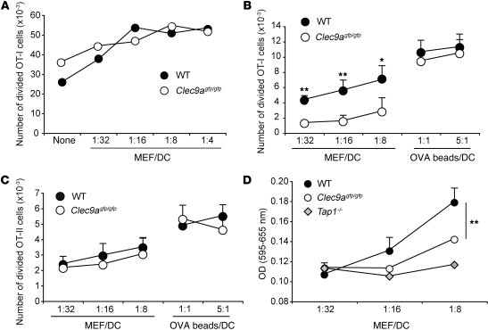
DNGR-1 (CLEC9A) is a receptor for necrotic cells required by DCs to cross-prime CTLs against dead cell antigens in mice. It is currently unknown how DNGR-1 couples dead cell recognition to cross-priming. Here we found that DNGR-1 did not mediate DC activation by dead cells but rather diverted necrotic cell cargo into a recycling endosomal compartment, favoring cross-presentation to CD8(+) T cells. DNGR-1 regulated cross-priming in non-infectious settings such as immunization with antigen-bearing dead cells, as well as in highly immunogenic situations such as infection with herpes simplex virus type 1. Together, these results suggest that DNGR-1 is a dedicated receptor for cross-presentation of cell-associated antigens. Our work thus underscores the importance of cross-priming in immunity and indicates that antigenicity and adjuvanticity can be decoded by distinct innate immune receptors. The identification of specialized receptors that regulate antigenicity of virus-infected cells reveals determinants of antiviral immunity that might underlie the human response to infection and vaccination.


Figures

Figure 1
Figure 1. DNGR-1 is required for cross-priming but not for cross-tolerance to dead cell–associated antigens.
(A) WT mice were left untreated or were injected with UV-treated H-2bm1 OVA-expressing MEFs i.v. One week later mice received B16-OVA cells i.v., and the number of tumors per lung was determined after 18 days. Data are representative of 2 independent experiments (n = 6/group, each dot represents 1 mouse). (B) WT (n = 16), DNGR-1–deficient (n = 11), or IAα–/– (n = 6) mice were injected with UV-treated H-2bm1 OVA–expressing MEFs, and the percentage of H2-Kb-OVA tetramer+ cells among CD8+ T cells in blood was determined 7 days after injection. Data were pooled from 2 independent experiments; each dot represents 1 mouse. (C) WT or DNGR-1–deficient mice received CFSE-labeled CD45.1+ OT-I cells, followed by immunization with UV-treated OVA-MEFs 1 day later. Spleens were analyzed 5 days after immunization for OT-I proliferation. CFSE dilution (left panel) and total number of divided cells (right panel) are shown; each dot represents 1 mouse. (D) DNGR-1–sufficient or DNGR-1–deficient RIP-mOVA mice received CFSE-labeled CD45.1+ OT-I cells. Renal LNs were analyzed 3 days after injection for OT-I proliferation quantified by CFSE dilution (left panel) and total number of divided cells (right panel); each dot represents 1 mouse. *P < 0.05, **P < 0.01, ***P < 0.001, unpaired Student’s t test and 1-way ANOVA (Tukey’s post-test).
Figure 2
Figure 2. DNGR-1 does not behave as a myeloid activatory receptor.
BM cells from Dectin-1–deficient mice were retrovirally transduced with a vector encoding for Dectin-1 or for a chimeric receptor (Chimera) bearing the cytoplasmic tail of DNGR-1 (WT) or 3 mutant versions thereof (Y7F, A3D, and I6G) fused to the transmembrane, stalk region, and CTLD of Dectin-1. All constructs were followed by an IRES-GFP sequence, which allowed sorting based on GFP levels to normalize for Dectin-1 or chimera surface expression. (A) Schematic representation of the chimeric DNGR-1/Dectin-1 receptors. Asterisks denote the mutated residues. (B) Expression of Dectin-1 or the different chimeras by GFP+ GMCSF-BMDCs purified by cell sorting as determined by staining with an anti–Dectin-1 antibody before overnight stimulation. (C) Purified GFP+ DCs as in B were cultured overnight in the presence of increasing amounts of curdlan (0, 30, and 100 μg/ml). Concentrations of TNF-α, IL-2, and IL-10 in the supernatant after culture are shown. Results are mean ± sem and representative of at least 3 independent experiments. ND, not detected.
Figure 3
Figure 3. DNGR-1 regulates MHC class I cross-presentation but not MHC class II presentation of dead cell–associated antigens.
(A) The adjuvanticity of dead cells is independent of DNGR-1. Purified CD8α+-like Flt3L-BMDCs from WT or DNGR-1–deficient (Clec9agfp/gfp) mice were pulsed with 1 pM SIINFEKL peptide, washed, and cultured with UV-treated H-2bm1 MEFs and CFSE-labeled OT-I T cells. OT-I proliferation was quantified after 3 days. (B and C) DNGR-1 regulates cross-presentation of dead cell–associated antigens. Purified CD8α+-like Flt3L-BMDCs from WT or DNGR-1–deficient mice were cultured with UV-treated OVA-MEFs or latex beads coated with OVA (OVA beads). Proliferation of CFSE-labeled OT-I (B) or OT-II (C) T cells was quantified on day 3 or 5 (respectively) of coculture with DCs. (D) Purified CD8α+-like Flt3L-BMDCs from WT, DNGR-1–deficient, or Tap1–/– mice were cultured with UV-treated OVA-MEFs and B3Z cells. NFAT reporter activity in B3Z cells was measured using a colorimetric assay after 24 hours of coculture. Results are mean ± sd and representative of at least 3 independent experiments. *P < 0.05, **P < 0.01, unpaired Student’s t test.
Figure 4
Figure 4. DNGR-1 diverts cargo to the early/recycling endosomal pool.
(A) Purified splenic CD8α+ DCs from WT mice were cultured for 30 minutes (EEA-1) or for 90 minutes (Rab5a and Rab27a) with Alexa Fluor 488–coupled anti–DNGR-1 Ab. Cells were fixed and stained for confocal microscopy. Distribution of internalized anti–DNGR-1 Ab+ vesicles relative to EEA-1+, Rab5a+, or Rab27a+ vesicles was quantified as percentage of vesicles positive for anti–DNGR-1 antibody also positive for endosomal markers in 15 cells per condition. (B and C) Purified CD8α+-like Flt3L-BMDCs from WT or DNGR-1–deficient mice were cultured with UV-treated Alexa Fluor 647–labeled H-2bm1 MEFs. Cells were fixed after 60 or 240 minutes and stained for confocal microscopy. Distribution of cargo derived from dead cells relative to Rab5a (B) and Rab11 (C) was quantified as percentage of total vesicles positive for endosomal markers containing dead cell cargo in 15 cells per condition. Scale bars: 5 μm. Results are representative of 2 independent experiments. *P < 0.05, **P < 0.01, unpaired Student’s t test.
Figure 5
Figure 5. SFV-OVA–infected cells induce TLR3-dependent DC activation and DNGR-1–dependent cross-presentation.
Purified CD8α+-like Flt3L-BMDCs from WT, DNGR-1–deficient, or Tlr3–/– mice were cultured with SFV-OVA–infected H-2bm1 MEFs (infected cells) at the indicated ratios in the absence (A) or presence (B) of CFSE-labeled OT-I T cells. (A) Concentration of IL-6 and TNF-α in the supernatant (upper panels) and MFI of CD86 and CD40 surface expression (lower panels) after overnight culture. As a positive control, DCs were cultured in the presence of CpG (500 ng/ml). Results are mean ± sem and representative of 2 independent experiments. (B) OT-I proliferation was quantified after 3 days. Results are mean ± sem and representative of at least 3 independent experiments. *P < 0.05, **P < 0.01, unpaired Student’s t test.
Figure 6
Figure 6. DNGR-1 contributes to the CTL response to HSV-1 lung infection.
WT, DNGR-1–deficient, or Batf3–/– mice were infected with HSV-1 i.n. Mediastinal LNs were harvested 7 days later, and CTL responses were measured directly ex vivo or after in vitro restimulation. (A) Left panel: H-2Kb-gB pentamer+ cells as a percentage of CD8+CD44+ T lymphocytes. Right panel: Number of H-2Kb-gB pentamer+CD8+CD44+ T cells. (B) IFN-γ+ cells as a percentage of CD8+CD44+ T lymphocytes after restimulation with gB498–505 peptide for 6 hours. Data were pooled from 3 independent experiments, with each dot representing 1 mouse; naive WT, n = 5; WT, n = 13; DNGR-1–deficient, n = 13, Batf3–/–, n = 8 mice. *P < 0.05, **P < 0.01, ***P < 0.001, 1-way ANOVA (Tukey’s post-test).

References

    1. Steinman RM. Lasker Basic Medical Research Award. Dendritic cells: versatile controllers of the immune system. Nat Med. 2007;13(10):1155–1159. doi: 10.1038/nm1643. - DOI - PubMed
    1. Steinman RM. Dendritic cells in vivo: a key target for a new vaccine science. Immunity. 2008;29(3):319–324. doi: 10.1016/j.immuni.2008.08.001. - DOI - PubMed
    1. Villadangos JA, Schnorrer P. Intrinsic and cooperative antigen-presenting functions of dendritic-cell subsets in vivo. Nat Rev Immunol. 2007;7(7):543–555. doi: 10.1038/nri2103. - DOI - PubMed
    1. Heath WR, Carbone FR. Dendritic cell subsets in primary and secondary T cell responses at body surfaces. Nat Immunol. 2009;10(12):1237–1244. doi: 10.1038/ni.1822. - DOI - PubMed
    1. Merad M, Manz MG. Dendritic cell homeostasis. Blood. 2009;113(15):3418–3427. doi: 10.1182/blood-2008-12-180646. - DOI - PMC - PubMed

Publication types

MeSH terms