. 2012 Aug;40(14):6808-20.
doi: 10.1093/nar/gks293. Epub 2012 Apr 13.

Argonaute identity defines the length of mature mammalian microRNAs

Affiliations

Argonaute identity defines the length of mature mammalian microRNAs

Prasanna Kumar Juvvuna et al. Nucleic Acids Res. 2012 Aug.

Abstract

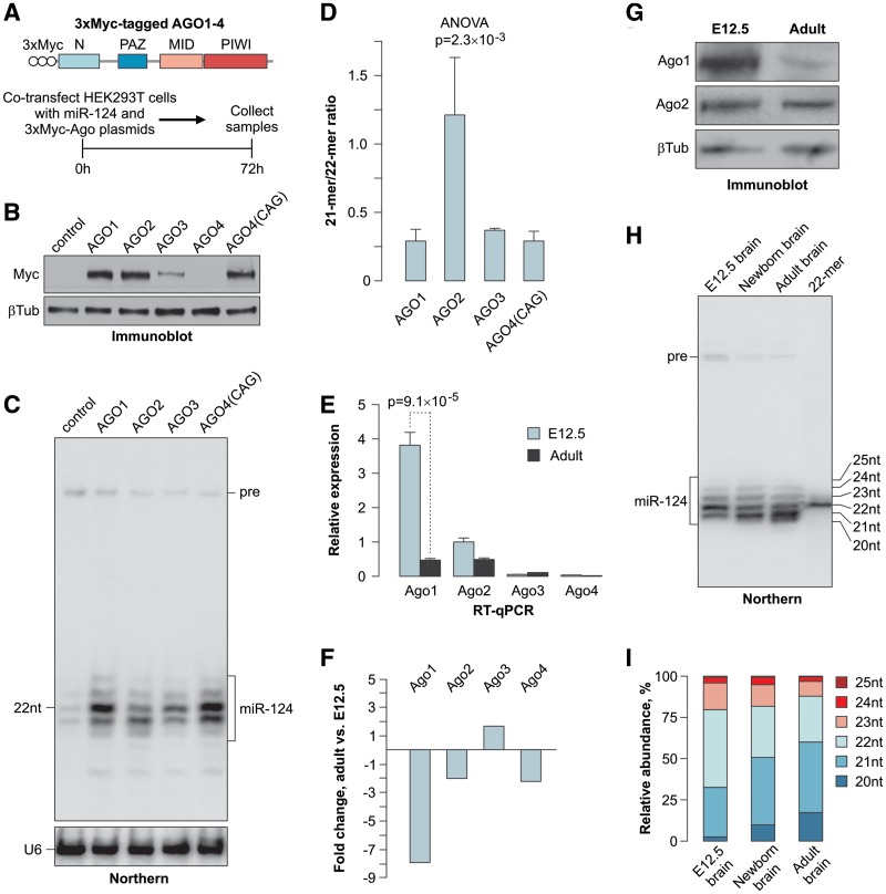
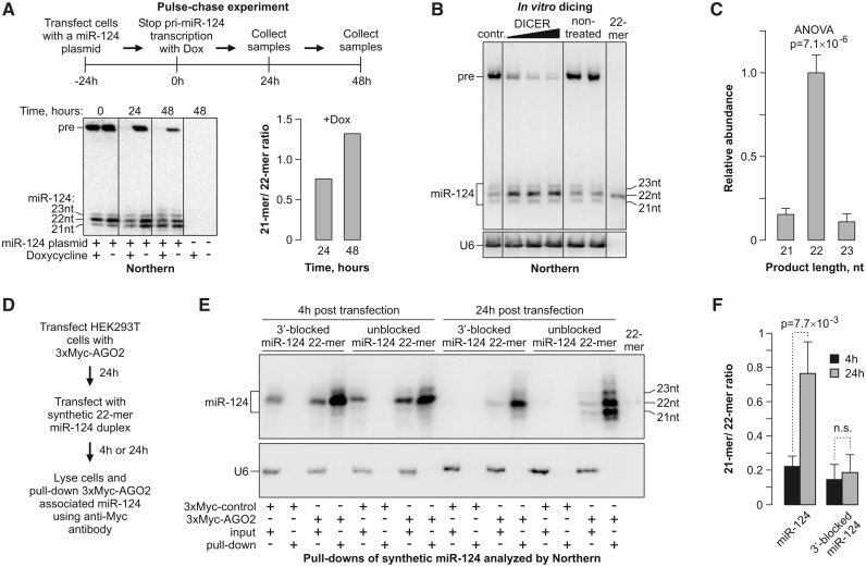
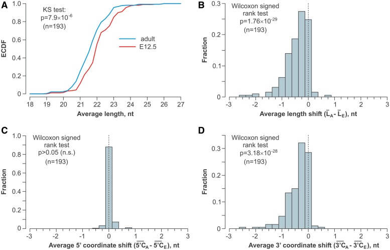
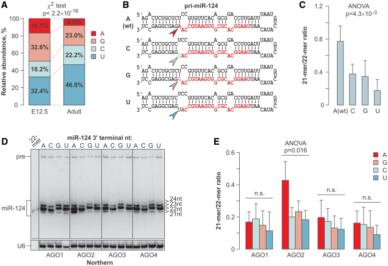
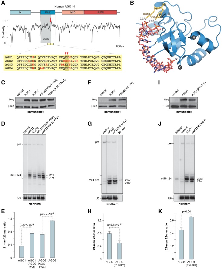
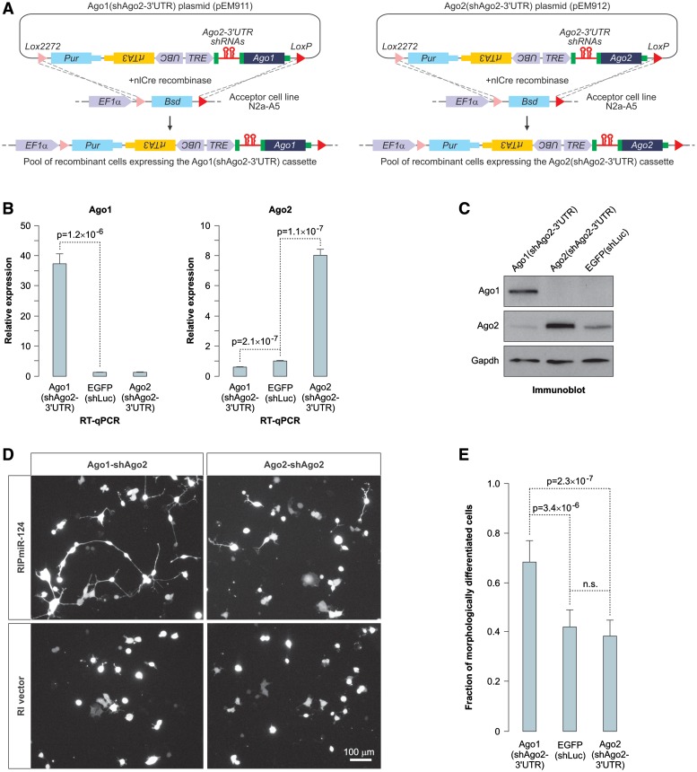
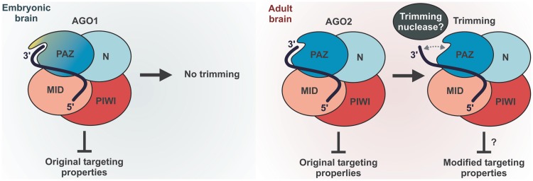
MicroRNAs (miRNAs) are 19- to 25-nt-long non-coding RNAs that regulate gene expression by base-pairing with target mRNAs and reducing their stability or translational efficiency. Mammalian miRNAs function in association with four closely related Argonaute proteins, AGO1-4. All four proteins contain the PAZ and the MID domains interacting with the miRNA 3' and 5' termini, respectively, as well as the PIWI domain comprising an mRNA 'slicing' activity in the case of AGO2 but not AGO1, AGO3 and AGO4. However, the slicing mode of the miRNA-programmed AGO2 is rarely realized in vivo and the four Argonautes are thought to play largely overlapping roles in the mammalian miRNA pathway. Here, we show that the average length of many miRNAs is diminished during nervous system development as a result of progressive shortening of the miRNA 3' ends. We link this modification with an increase in the fractional abundance of Ago2 in the adult brain and identify a specific structural motif within the PAZ domain that enables efficient trimming of miRNAs associated with this but not the other three Argonautes. Taken together, our data suggest that mammalian Argonautes may define the length and possibly biological activity of mature mammalian miRNAs in a developmentally controlled manner.


Figures

Figure 1.
Figure 1.
Mammalian Argonautes modulate the mature miRNA length. (A) Top, diagram of the 3xMyc-AGO constructs and bottom, workflow of the 3xMyc AGO/miR-124 co-expression experiment. (B) Immunoblot analysis of the 3xMyc-tagged AGO1–4 proteins expressed from a CMV promoter and AGO4 expressed from a CAG promoter using a Myc tag-specific antibody. β-tubulin is a lane loading control. (C) Northern-blot analysis of miR-124 co-expressed with the corresponding 3xMyc AGO proteins or a control plasmid. The positions of the mature miR-124 and the pre-miR-124 precursor (pre) are marked on the right. U6 snRNA specific probe is used as a loading control. (D) Quantification of the results in (C) showing a significantly increased ratio between the 21- and 22-mer in cells over-expressing 3xMyc AGO2 as compared to the other three Argonautes. Data are averaged from three independent transfection experiments ±SD. (E) RT-qPCR analysis of the Argonaute expression in E12.5 and adult (P60) mouse brains. Data were averaged from three biological replicates ±SD and analyzed by the t test. (F) Quantification of the data in (E) showing a dramatic decrease in the Ago1 expression in the adult brain. Negative fold changes correspond to down-regulation in the adult brain and positive fold changes correspond to up-regulation in the adult brain. (G) Immunoblot analysis showing a relatively uniform expression of Ago2 in the E12.5 and adult mouse brains and a reduction in the Ago1 protein levels during mouse brain development. β-tubulin is a loading control. (H) Northern-blot analysis of RNAs from E12.5, newborn (P0) and adult (P60) mouse brains showing progressive shortening of mature miR-124 during development. (I) Quantification of the data in (H) showing a change in the percentage of the corresponding miR-124 isoforms (20–25 nt) as a function of development.
Figure 2.
Figure 2.
miRNA length is reduced following its recruitment to AGO2. (A) Pulse-chase experiment showing that the mature miR-124 length is reduced after the completion of the Drosha- and Dicer-dependent processing steps. Top, the experimental workflow; bottom left, Northern-blot analysis of the corresponding RNA samples; bottom right, quantification of the Northern-blot data showing an increase in the 21- to 22-mer ratio in samples from collected 24 and 48 h after halting the pri-miR-124 transcription by Dox. (B) RNA from miR-124-expressing HEK293T cells in (A) at the time point 0 was incubated in vitro with increasing amounts of purified human Dicer and the reaction products were analyzed by Northern blotting. (C) Quantification of the newly formed products in (B) showing that Dicer generates predominantly 22-mer species. Data are averaged from the three Dicer lanes in (B) ±SD. (D–F) AGO2-loaded synthetic 22-mers are efficiently trimmed unless protected at the 3 end. (D) Outline of the experiment. (E) HEK293T cells expressing 3xMyc-AGO2 were transfected with synthetic siRNA-like duplexes containing either an unmodified 22-mer miR-124 or 22-mer protected at the 3 end with a 2-OMe group. The AGO2/miR-124 complexes were pulled down 4- and 24-h post-transfection and analyzed by Northern blotting. (F) Quantifications of the data in (E) confirming efficient trimming of the unmodified but not 3-protected 22-mers loaded into AGO2. Data are averaged from three independent experiments ±SD.
Figure 3.
Figure 3.
Widespread shortening of the miRNA 3 ends during brain development. Statistical analyses of deep sequencing data for 193 microRNAs expressed in the E12.5 and the adult mouse brain and containing no non-templated nucleotides. (A) The overall length distribution of miRNAs expressed in the adult brain is shifted toward shorter species as compared to E12.5. (B) Average lengths of specific miRNAs tend to decrease in the adult brain as compared to E12.5. (C) The miRNA 5 end coordinates do not differ significantly between the two brain samples. (D) The 3 end coordinates of the adult miRNAs are diminished significantly as compared to E12.5.
Figure 4.
Figure 4.
3-Terminal nucleotide preferences of the trimming reaction. (A) The usage of the 3-terminal genome-encoded nucleotides is relatively uniform in the E12.5 miRNAs, whereas it becomes significantly depleted in A’s and enriched in U’s in the adult sample. (B) Diagram of the miR-124 expression constructs genetically modified to introduce all 4 nt (arrowheads) at the 3-terminal position of the mature miRNA (colored sequence). (C) Of the four miR-124 variants described in (B), A3 is trimmed most efficiently and U3 is trimmed least efficiently in the HEK293T cells. (D) The four 3-terminal miR-124 variants were co-expressed in HEK293T cells with the AGO1–4 and the trimming efficiency was analyzed by Northern blotting 48-h post-transfection. (E) Quantification of the data in (D).
Figure 5.
Figure 5.
Structure of the Argonaute PAZ domain modulates miRNA trimming efficiency. (A) Human Argonaute sequence similarity plot. The diagram on the top indicates the positions of the N, PAZ, MID and PIWI domains. The divergent sequence element within the PAZ domain is indicated by the arrow and the sequence swapped between AGO1 and AGO2 in (C–E) is highlighted in gray. A fragment of the PAZ sequence alignment is presented at the bottom with non-conserved amino acid residues shown in red. The position of the AGO1 K313 and Y314 residues is marked by the arrowheads. (B) Crystal structure of the AGO1 PAZ domain associated with a small RNA [1SI3; (14)] showing hydrogen bonds between the K313 and Y314 side chains and the 3 penultimate and ultimate nucleoside triphosphates. (C, F and I) Immunoblot analyses showing comparable expression of the wild-type AGO1 and AGO2, (C) their reciprocal PAZ domain-swapped mutants, (F) AGO2(RH > KY) mutant and (I) AGO1(KY > RH) mutant in HEK293T cells. (D, G and J) Northern-blot analyses of miR-124 co-expressed with the corresponding Argonaute proteins in HEK293T cells. Cells were harvested at (D) 72 hours or (G and J) 48-h post-transfection. (E, H and K) Quantification of the data in (D, G and J) showing the effect of the PAZ domain structure on the miRNA trimming efficiency. Data were averaged from three independent transfection experiments ±SD and compared using the t test.
Figure 6.
Figure 6.
AGO1 stimulates miR-124-induced neuronal differentiation of neuroblastoma cells. (A) Diagram of the HILO-RMCE procedure used to obtain pools of transgenic N2a cells expressing Dox-inducible Ago1(shAgo2-3-UTR) and Ago2(shAgo2-3-UTR) cassettes. (B and C) Transgenic N2a cells generated as described in (A) or cells containing a Dox-inducible EGFP(shLuc) control transgene were treated with Dox for 72 h and the Ago1 and Ago2 expression was analyzed by (B) RT-qPCR or (C) immunoblotting. Data in (B) were averaged from six independent amplification experiments ±SD and compared using t test. (D) Ago1(shAgo2-3-UTR) and Ago2(shAgo2-3-UTR) cells pre-treated with Dox for 72 h were transiently transfected with the miR-124 expression plasmid RIP-miR-124 additionally encoding the dsRed2 fluorescent marker or the corresponding control vector (RI vector) and the transfected cell morphology was examined 36-h post-transfection using dsRed2 fluorescence. Note that the expression of RIP-miR-124 leads to the appearance of a larger number of neuron-like cells containing long processes in the Ago1(shAgo2-3-UTR) cells as compared to the Ago2(shAgo2-3-UTR) cells. No differentiated cells are obvious in the RI vector-transfected cultures. (E) Quantification of the RIPmiR-124-induced morphological differentiation in Ago1(shAgo2-3-UTR), Ago2(shAgo2-3-UTR) and EGFP(shLuc) cells. Shown are fractions of cells containing at least one primary process longer than the average diameter of the cell soma. Ten randomly selected fields were imaged for each sample and the data were averaged ±SD and compared using the t test.
Figure 7.
Figure 7.
Model outlining the role of mammalian Argonaute identity in miRNA trimming.

Similar articles

Cited by

References

    1. Fabian MR, Sonenberg N, Filipowicz W. Regulation of mRNA translation and stability by microRNAs. Annu. Rev. Biochem. 2010;79:351–379. - PubMed
    1. Kim VN, Han J, Siomi MC. Biogenesis of small RNAs in animals. Nat. Rev. Mol. Cell. Biol. 2009;10:126–139. - PubMed
    1. Bartel DP. MicroRNAs: target recognition and regulatory functions. Cell. 2009;136:215–233. - PMC - PubMed
    1. Ivey KN, Srivastava D. MicroRNAs as regulators of differentiation and cell fate decisions. Cell Stem Cell. 2010;7:36–41. - PubMed
    1. Fineberg SK, Kosik KS, Davidson BL. MicroRNAs potentiate neural development. Neuron. 2009;64:303–309. - PubMed

Publication types