

p53 N-terminal phosphorylation: a defining layer of complex regulation
- PMID: 22505655
- PMCID: PMC3499055
- DOI: 10.1093/carcin/bgs145
p53 N-terminal phosphorylation: a defining layer of complex regulation
Abstract
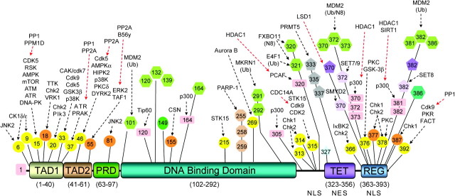
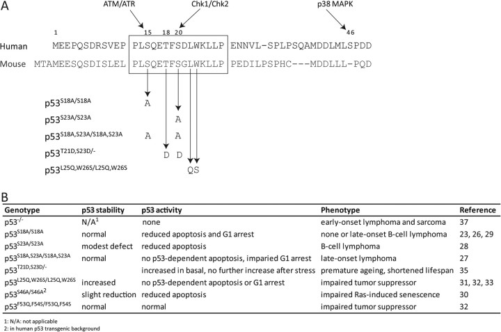
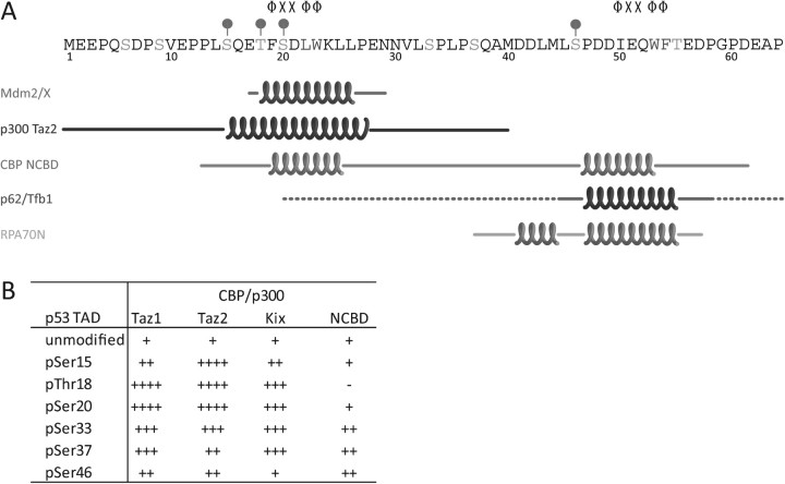
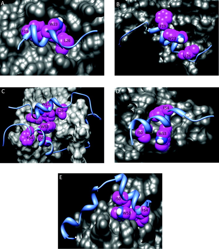
The p53 tumor suppressor is a critical component of the cellular response to stress. As it can inhibit cell growth, p53 is mutated or functionally inactivated in most tumors. A multitude of protein-protein interactions with transcriptional cofactors are central to p53-dependent responses. In its activated state, p53 is extensively modified in both the N- and C-terminal regions of the protein. These modifications, especially phosphorylation of serine and threonine residues in the N-terminal transactivation domain, affect p53 stability and activity by modulating the affinity of protein-protein interactions. Here, we review recent findings from in vitro and in vivo studies on the role of p53 N-terminal phosphorylation. These modifications can either positively or negatively affect p53 and add a second layer of complex regulation to the divergent interactions of the p53 transactivation domain.
Figures
Similar articles
-
Signaling to p53: breaking the posttranslational modification code.Pathol Biol (Paris). 2000 Apr;48(3):227-45. Pathol Biol (Paris). 2000. PMID: 10858956 Review.
-
Phosphorylation Regulates the Bound Structure of an Intrinsically Disordered Protein: The p53-TAZ2 Case.PLoS One. 2016 Jan 7;11(1):e0144284. doi: 10.1371/journal.pone.0144284. eCollection 2016. PLoS One. 2016. PMID: 26742101 Free PMC article.
-
Serine15 phosphorylation stimulates p53 transactivation but does not directly influence interaction with HDM2.EMBO J. 1999 Dec 15;18(24):7002-10. doi: 10.1093/emboj/18.24.7002. EMBO J. 1999. PMID: 10601022 Free PMC article.
-
Activation of p53 by oxidative stress involves platelet-derived growth factor-beta receptor-mediated ataxia telangiectasia mutated (ATM) kinase activation.J Biol Chem. 2003 Oct 10;278(41):39527-33. doi: 10.1074/jbc.M304423200. Epub 2003 Jul 30. J Biol Chem. 2003. PMID: 12890678
-
Mechanisms of transcriptional regulation by p53.Cell Death Differ. 2018 Jan;25(1):133-143. doi: 10.1038/cdd.2017.174. Epub 2017 Nov 10. Cell Death Differ. 2018. PMID: 29125602 Free PMC article. Review.
Cited by
-
C16-ceramide is a natural regulatory ligand of p53 in cellular stress response.Nat Commun. 2018 Oct 8;9(1):4149. doi: 10.1038/s41467-018-06650-y. Nat Commun. 2018. PMID: 30297838 Free PMC article.
-
Recognition of the disordered p53 transactivation domain by the transcriptional adapter zinc finger domains of CREB-binding protein.Proc Natl Acad Sci U S A. 2016 Mar 29;113(13):E1853-62. doi: 10.1073/pnas.1602487113. Epub 2016 Mar 14. Proc Natl Acad Sci U S A. 2016. PMID: 26976603 Free PMC article.
-
CK2 phosphorylates and inhibits TAp73 tumor suppressor function to promote expression of cancer stem cell genes and phenotype in head and neck cancer.Neoplasia. 2014 Oct 23;16(10):789-800. doi: 10.1016/j.neo.2014.08.014. eCollection 2014 Oct. Neoplasia. 2014. PMID: 25379016 Free PMC article.
-
Targeting 7KCh-Induced Cell Death Response Mediated by p38, P2X7 and GSDME in Retinal Pigment Epithelium Cells with Sterculic Acid.Pharmaceutics. 2023 Nov 5;15(11):2590. doi: 10.3390/pharmaceutics15112590. Pharmaceutics. 2023. PMID: 38004569 Free PMC article.
-
Reactivation of Epstein-Barr Virus by HIF-1α Requires p53.J Virol. 2020 Aug 31;94(18):e00722-20. doi: 10.1128/JVI.00722-20. Print 2020 Aug 31. J Virol. 2020. PMID: 32641480 Free PMC article.
References
-
- Petitjean A, et al. Impact of mutant p53 functional properties on TP53 mutation patterns and tumor phenotype: lessons from recent developments in the IARC TP53 database. Hum. Mutat. 2007;28:622–629. - PubMed
-
- Goh AM, et al. The role of mutant p53 in human cancer. J. Pathol. 2011;223:116–126. - PubMed
-
- Woods DB, et al. Regulation of p53 function. Exp. Cell Res. 2001;264:56–66. - PubMed
-
- El-Deiry WS, et al. Definition of a consensus binding site for p53. Nat. Genet. 1992;1:45–49. - PubMed
Publication types
MeSH terms
Substances
Grants and funding
LinkOut - more resources
Full Text Sources
Other Literature Sources
Molecular Biology Databases
Research Materials
Miscellaneous
