

Molecular cloning and characterization of first organic matrix protein from sclerites of red coral, Corallium rubrum
- PMID: 22505718
- PMCID: PMC3365975
- DOI: 10.1074/jbc.M112.352005
Molecular cloning and characterization of first organic matrix protein from sclerites of red coral, Corallium rubrum
Abstract
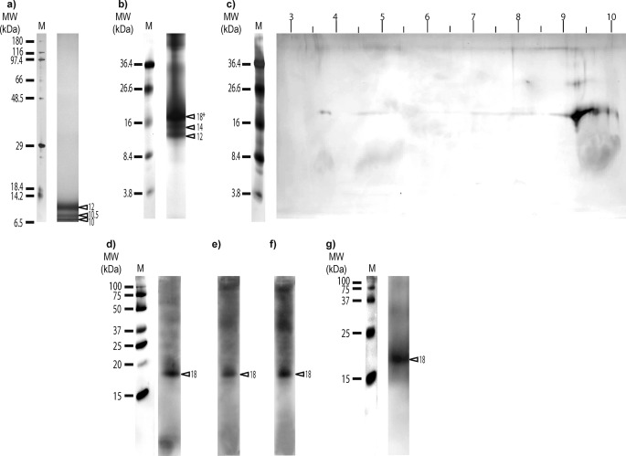
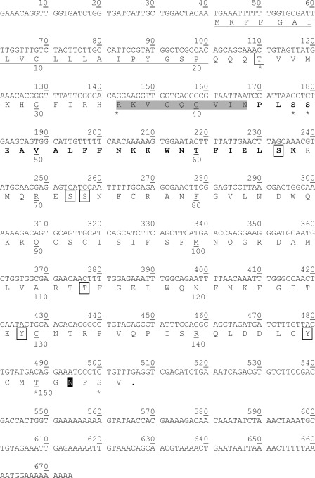
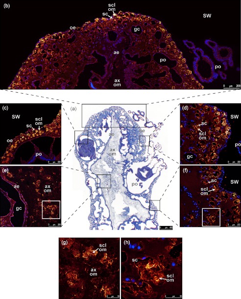
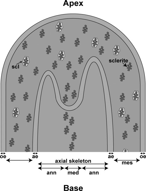
We report here for the first time the isolation and characterization of a protein from the organic matrix (OM) of the sclerites of the alcyonarian, Corallium rubrum. This protein named scleritin is one of the predominant proteins extracted from the EDTA-soluble fraction of the OM. The entire open reading frame (ORF) was obtained by comparing amino acid sequences from de novo mass spectrometry and Edman degradation with an expressed sequence tag library dataset of C. rubrum. Scleritin is a secreted basic phosphorylated protein which exhibits a short amino acid sequence of 135 amino acids and a signal peptide of 20 amino acids. From specific antibodies raised against peptide sequences of scleritin, we obtained immunolabeling of scleroblasts and OM of the sclerites which provides information on the biomineralization pathway in C. rubrum.
Figures
Similar articles
-
The skeletome of the red coral Corallium rubrum indicates an independent evolution of biomineralization process in octocorals.BMC Ecol Evol. 2021 Jan 11;21(1):1. doi: 10.1186/s12862-020-01734-0. BMC Ecol Evol. 2021. PMID: 33514311 Free PMC article.
-
Molecular cloning of a cDNA encoding a soluble protein in the coral exoskeleton.Biochem Biophys Res Commun. 2003 Apr 25;304(1):11-7. doi: 10.1016/s0006-291x(03)00527-8. Biochem Biophys Res Commun. 2003. PMID: 12705876
-
Comparative analysis of the soluble organic matrix of axial skeleton and sclerites of Corallium rubrum: insights for biomineralization.Comp Biochem Physiol B Biochem Mol Biol. 2011 May;159(1):40-8. doi: 10.1016/j.cbpb.2011.01.007. Epub 2011 Jan 28. Comp Biochem Physiol B Biochem Mol Biol. 2011. PMID: 21281736
-
Complete mitochondrial genomes of the Japanese pink coral (Corallium elatius) and the Mediterranean red coral (Corallium rubrum): a reevaluation of the phylogeny of the family Coralliidae based on molecular data.Comp Biochem Physiol Part D Genomics Proteomics. 2013 Sep;8(3):209-19. doi: 10.1016/j.cbd.2013.05.003. Epub 2013 Jun 2. Comp Biochem Physiol Part D Genomics Proteomics. 2013. PMID: 23792378
-
An alternative and effective method for extracting skeletal organic matrix adapted to the red coral Corallium rubrum.Biol Open. 2022 Oct 15;11(10):bio059536. doi: 10.1242/bio.059536. Epub 2022 Oct 14. Biol Open. 2022. PMID: 36178163 Free PMC article.
Cited by
-
Activation Profile Analysis of CruCA4, an α-Carbonic Anhydrase Involved in Skeleton Formation of the Mediterranean Red Coral, Corallium rubrum.Molecules. 2017 Dec 28;23(1):66. doi: 10.3390/molecules23010066. Molecules. 2017. PMID: 29283417 Free PMC article.
-
Comparative Proteomics of Octocoral and Scleractinian Skeletomes and the Evolution of Coral Calcification.Genome Biol Evol. 2020 Sep 1;12(9):1623-1635. doi: 10.1093/gbe/evaa162. Genome Biol Evol. 2020. PMID: 32761183 Free PMC article.
-
The skeletal proteome of the coral Acropora millepora: the evolution of calcification by co-option and domain shuffling.Mol Biol Evol. 2013 Sep;30(9):2099-112. doi: 10.1093/molbev/mst109. Epub 2013 Jun 12. Mol Biol Evol. 2013. PMID: 23765379 Free PMC article.
-
Carbonic Anhydrases in Cnidarians: Novel Perspectives from the Octocorallian Corallium rubrum.PLoS One. 2016 Aug 11;11(8):e0160368. doi: 10.1371/journal.pone.0160368. eCollection 2016. PLoS One. 2016. PMID: 27513959 Free PMC article.
-
The skeleton of the staghorn coral Acropora millepora: molecular and structural characterization.PLoS One. 2014 Jun 3;9(6):e97454. doi: 10.1371/journal.pone.0097454. eCollection 2014. PLoS One. 2014. PMID: 24893046 Free PMC article.
References
-
- Estroff L. A. (2008) Introduction: biomineralization. Chem. Rev. 108, 4329–4331 - PubMed
-
- Lowenstam H. A., Weiner S. (1989) On Biomineralization, pp. 7–16, Oxford University Press, New York
-
- Marin F., Luquet G., Marie B., Medakovic D. (2008) Molluscan shell proteins: primary structure, origin, and evolution. Curr. Top. Dev. Biol. 80, 209–276 - PubMed
-
- Mann S. (2001) Biomineralization: Principles and Concepts in Bioinorganic Materials Chemistry (Compton R. G., Davies S. G., Evans J., eds) pp. 6–23, Oxford University Press, New York
-
- Dubois P., Chen C. P. (1989) in Echinoderm Studies (Jangoux M., Lawrence J. M., eds) pp. 109–178, Rotterdam
Publication types
MeSH terms
Substances
Associated data
- Actions
LinkOut - more resources
Full Text Sources
Other Literature Sources
