

Temporal and quantitative analysis of atherosclerotic lesions in diet-induced hypercholesterolemic rabbits
- PMID: 22505812
- PMCID: PMC3312324
- DOI: 10.1155/2012/506159
Temporal and quantitative analysis of atherosclerotic lesions in diet-induced hypercholesterolemic rabbits
Abstract
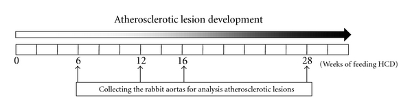
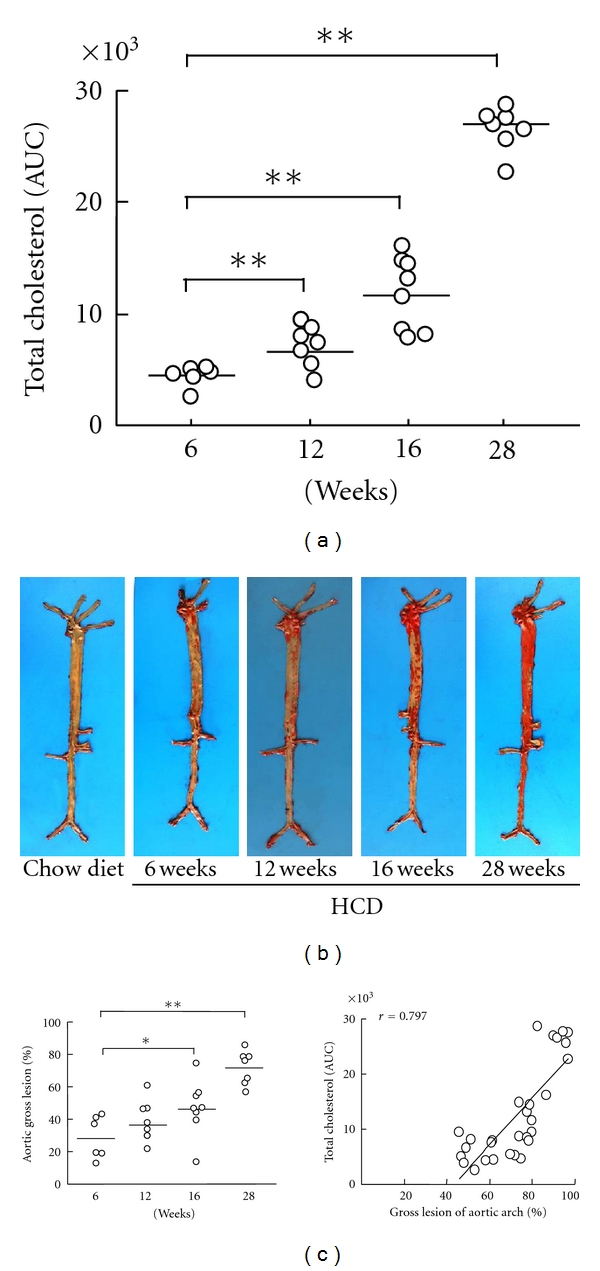
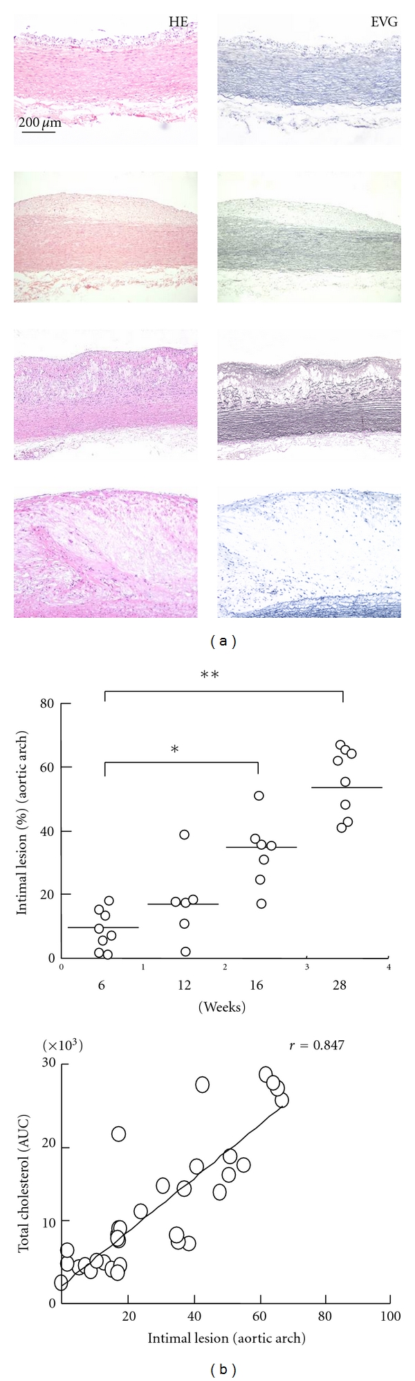
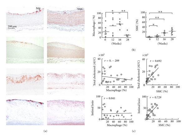
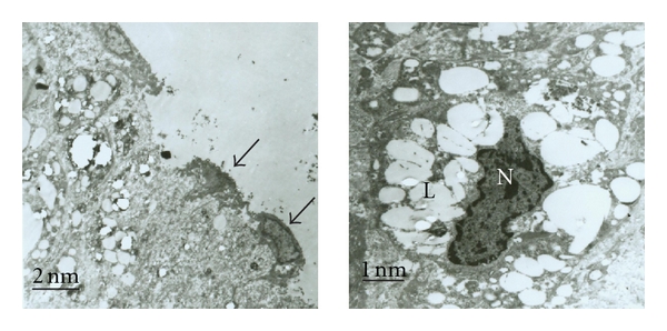
The diet-induced atherosclerotic rabbit is an ideal model for atherosclerosis study, but temporal changes in atherosclerotic development in hypercholesterolemic rabbits are poorly understood. Japanese white rabbits were fed a high-cholesterol diet to induce sustained hypercholesterolemia, and each group of 10-12 animals was then sacrificed at 6, 12, 16, or 28 weeks. The rabbit aortas were harvested, and the sizes of the gross and intima atherosclerotic lesions were quantified. The cellular component of macrophages (Mφs) and smooth muscle cells (SMCs) in aortic intimal lesions was also quantified by immunohistochemical staining, and the correlation between plasma cholesterol levels and the progress of atherosclerotic lesions was studied. The ultrastructure of the atherosclerotic lesions was observed by transmission electron microscopy (TEM). Widely variable atherosclerotic plaques were found from 6 weeks to 28 weeks, and the lesional progress was closely correlated with cholesterol exposure. Interestingly, a relatively reduced accumulation of Mφ, an increased numbers of SMCs, and a damaged endothelial layer were presented in advanced lesions. Moreover, SMCs were closely correlated with cholesterol exposure and lesional progress for the whole period. Cholesterol exposure directly determines atherosclerotic progress in a rabbit model, and the changes in the cellular component of advanced lesions may affect plaque stability in an atherosclerotic rabbit model.
Figures
Similar articles
-
Increased Hepatic Expression of Endothelial Lipase Inhibits Cholesterol Diet-Induced Hypercholesterolemia and Atherosclerosis in Transgenic Rabbits.Arterioscler Thromb Vasc Biol. 2017 Jul;37(7):1282-1289. doi: 10.1161/ATVBAHA.117.309139. Epub 2017 May 25. Arterioscler Thromb Vasc Biol. 2017. PMID: 28546217 Free PMC article.
-
C-reactive protein levels are associated with the progression of atherosclerotic lesions in rabbits.Histol Histopathol. 2012 Apr;27(4):529-35. doi: 10.14670/HH-27.529. Histol Histopathol. 2012. PMID: 22374730
-
Dietary cholesterol atherogenic changes in juvenile rabbits.Biol Pharm Bull. 2015;38(5):785-8. doi: 10.1248/bpb.b14-00775. Biol Pharm Bull. 2015. PMID: 25947925
-
Emerging Concepts of Vascular Cell Clonal Expansion in Atherosclerosis.Arterioscler Thromb Vasc Biol. 2022 Mar;42(3):e74-e84. doi: 10.1161/ATVBAHA.121.316093. Epub 2022 Feb 3. Arterioscler Thromb Vasc Biol. 2022. PMID: 35109671 Free PMC article. Review.
-
Early atherogenesis: new insights from new approaches.Curr Opin Lipidol. 2022 Oct 1;33(5):271-276. doi: 10.1097/MOL.0000000000000843. Epub 2022 Aug 12. Curr Opin Lipidol. 2022. PMID: 35979994 Free PMC article. Review.
Cited by
-
Fat-1 expression alleviates atherosclerosis in transgenic rabbits.J Cell Mol Med. 2022 Feb;26(4):1306-1314. doi: 10.1111/jcmm.17188. Epub 2022 Jan 18. J Cell Mol Med. 2022. PMID: 35040258 Free PMC article.
-
TdP Incidence in Methoxamine-Sensitized Rabbit Model Is Reduced With Age but Not Influenced by Hypercholesterolemia.Front Physiol. 2021 Jun 21;12:692921. doi: 10.3389/fphys.2021.692921. eCollection 2021. Front Physiol. 2021. PMID: 34234694 Free PMC article.
-
Antioxidant and anti-inflammatory effects of Marrubium alysson extracts in high cholesterol-fed rabbits.Saudi Pharm J. 2014 Nov;22(5):472-82. doi: 10.1016/j.jsps.2013.12.004. Epub 2013 Dec 21. Saudi Pharm J. 2014. PMID: 25473336 Free PMC article.
-
Increased expression of phosphorylated forms of heat-shock protein-27 and p38MAPK in macrophage-rich regions of fibro-fatty atherosclerotic lesions in the rabbit.Int J Exp Pathol. 2016 Feb;97(1):56-65. doi: 10.1111/iep.12167. Epub 2016 Feb 8. Int J Exp Pathol. 2016. PMID: 26853073 Free PMC article.
-
Infection with Porphyromonas gingivalis exacerbates endothelial injury in obese mice.PLoS One. 2014 Oct 21;9(10):e110519. doi: 10.1371/journal.pone.0110519. eCollection 2014. PLoS One. 2014. PMID: 25334003 Free PMC article.
References
-
- Libby P, Aikawa M. Stabilization of atherosclerotic plaques: new mechanisms and clinical targets. Nature Medicine. 2002;8(11):1257–1262. - PubMed
-
- Ross R. Atherosclerosis—an inflammatory disease. New England Journal of Medicine. 1999;340(2):115–126. - PubMed
-
- Steinberg D. Hypercholesterolemia and inflammation in atherogenesis: two sides of the same coin. Molecular Nutrition and Food Research. 2005;49(11):995–998. - PubMed
-
- Libby P. Changing concepts of atherogenesis. Journal of Internal Medicine. 2000;247(3):349–358. - PubMed
-
- Bennett MR. Apoptosis of vascular smooth muscle cells in vascular remodelling and atherosclerotic plaque rupture. Cardiovascular Research. 1999;41(2):361–368. - PubMed
Publication types
MeSH terms
Substances
LinkOut - more resources
Full Text Sources
Medical
