. 2012:2012:579141.
doi: 10.1155/2012/579141. Epub 2012 Mar 15.

Physical properties and biocompatibility of a core-sheath structure composite scaffold for bone tissue engineering in vitro

Affiliations

Physical properties and biocompatibility of a core-sheath structure composite scaffold for bone tissue engineering in vitro

Chuangjian Wang et al. J Biomed Biotechnol. 2012.

Abstract

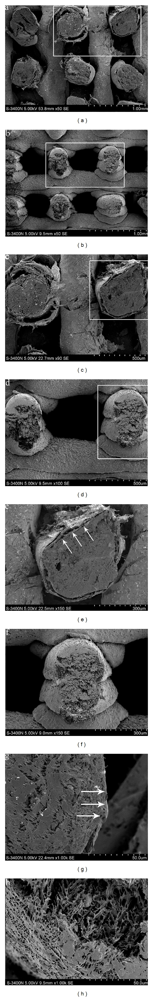
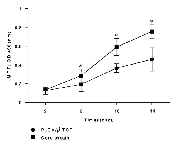
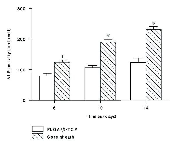
Scaffolds play a critical role in the practical realization of bone tissue engineering. The purpose of this study was to assess whether a core-sheath structure composite scaffold possesses admirable physical properties and biocompatibility in vitro. A novel scaffold composed of poly(lactic-co-glycolic acid)/β-tricalcium phosphate (PLGA/β-TCP) skeleton wrapped with Type I collagen via low-temperature deposition manufacturing (LDM) was prepared, and bone mesenchymal stem cells (BMSCs) were used to evaluate cell behavior on the scaffold. PLGA/β-TCP skeleton was chosen as the control group. Physical properties were evaluated by pority ratio, compressive strength, and Young's modulus. Scanning electron microscope (SEM) was used to study morphology of cells. Hydrophilicity was evaluated by water absorption ratio. Cell proliferation was tested by 3-(4,5-dimethylthiazol-2-yl)-2,5-diphenyl tetrazolium bromide assay (MTT). Osteogenic differentiation of BMSCs was evaluated by alkaline phosphates activity (ALP). The results indicated that physical properties of the novel scaffold were as good as those of the control group, hydrophilicity was observably better (P < 0.01) than that of control group, and abilities of proliferation and osteogenic differentiation of BMSCs on novel scaffold were significantly greater (P < 0.05) than those of control group, which suggests that the novel scaffold possesses preferable characteristics and have high value in bone tissue engineering.


Figures

Figure 1
Figure 1
(a and b) Morphological comparison observation by SEM of the core-sheath structure composite scaffold and the PLGA/β-TCP skeleton (×50,50). (c and e) Magnified view of the white rectangle frame from (a) (×90,150). Note: the arrows point to the collagen wrapped the PLGA/β-TCP. (d and f) Magnified view of the white rectangle frame from (b) (×100,150). (g) Morphological observation by SEM of the core-sheath structure composite scaffold (×1 k). Note: the arrows point to the interface of the PLGA/β-TCP and the collagen. (h) Morphological observation by SEM of the PLGA/β-TCP skeleton. (×1 k).
Figure 2
Figure 2
Cell proliferation comparisons on the core-sheath structure composite scaffold and the PLGA/β-TCP skeleton. Cell proliferation analysis by MTT methods. Each value and error bar represent the mean of triplicate samples and their standard deviation (*P < 0.05 compared with the PLGA/β-TCP scaffolds; n = 3).
Figure 3
Figure 3
(a) Morphological observation of BMSCs cultured on the core-sheath structure composite scaffold by SEM after 4 hours (×3.0 k). Note: the arrows point to the BMSCs adhered on the scaffold and the pseudopodium of the cells. (c and d) Morphological observation of BMSCs cultured on the core-sheath structure composite scaffold by SEM after 2 days (×500,1000). Note: the arrows point to the BMSCs stretched onto the scaffold. (g and h) Morphological observation of BMSCs cultured on the core-sheath structure composite scaffold by SEM after 6 days (×300,1000). Note: the arrows point to the BMSCs. (k, l, m, and n) Morphological observation of BMSCs cultured on the core-sheath structure composite scaffold by SEM after 14 days (×500,1000). Note: (k and l) the arrows point to the BMSCs and secreted ECM by BMSCs. (m and n) the arrows point to the BMSCs growed into the holes of the scaffold. (b) Morphological observation of BMSCs cultured on the PLGA/β-TCP skeleton after 4 hours (×3.0 k). (e and f) Morphological observation of BMSCs cultured on the PLGA/β-TCP skeleton after 2 days (×500,1000). Note: the arrows point to the BMSCs. (I and j) Morphological observation of BMSCs cultured on the PLGA/β-TCP skeleton after 6 days (×500,1000). Note: the arrows point to the BMSCs. (o and p) Morphological observation of BMSCs cultured on the PLGA/β-TCP skeleton after 14 days (×400,1000). Note: the arrows point to the BMSCs.
Figure 4
Figure 4
ALP activity comparisons on the core-sheath structure composite scaffold and the PLGA/β-TCP skeleton. Each value and error bar represent the mean of triplicate samples and their standard deviation (*P < 0.05 compared with the PLGA/β-TCP scaffolds; n = 3).

References

    1. Amstein CF, Hartman PA. Adaptation of plastic surfaces for tissue culture by glow discharge. Journal of Clinical Microbiology. 1975;2(1):46–54. - PMC - PubMed
    1. Shi S, Wang XH, Guo G, Fan M, Huang MJ, Qian ZY. Preparation and characterization of microporous poly(D,L-lactic acid) film for tissue engineering scaffold. International Journal of Nanomedicine. 2010;5(1):1049–1055. - PMC - PubMed
    1. Angwarawong T, Dubas ST, Arksornnukit M, Pavasant P. Differentiation of MC3T3-E1 on poly(4-styrenesulfonic acid-co-maleic acid) sodium salt-coated films. Dental Materials Journal. 2011;30(2):158–169. - PubMed
    1. Gao C, Hu X, Hong Y, Guan J, Shen J. Photografting of poly(hydroxylethyl acrylate) onto porous polyurethane scaffolds to improve their endothelial cell compatibility. Journal of Biomaterials Science, Polymer Edition. 2003;14(9):937–950. - PubMed
    1. Gao J, Niklason L, Langer R. Surface hydrolysis of poly(glycolic acid) meshes increases the seeding density of vascular smooth muscle cells. Journal of Biomedical Materials Research. 1998;42(3):417–424. - PubMed

MeSH terms

LinkOut - more resources