Efficiency of manual scanning in recovering rare cellular events identified by fluorescence in situ hybridization: simulation of the detection of fetal cells in maternal blood
- PMID: 22505816
- PMCID: PMC3312578
- DOI: 10.1155/2012/610856
Efficiency of manual scanning in recovering rare cellular events identified by fluorescence in situ hybridization: simulation of the detection of fetal cells in maternal blood
Abstract
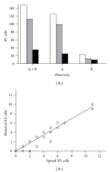
Fluorescence in situ hybridization (FISH) and manual scanning is a widely used strategy for retrieving rare cellular events such as fetal cells in maternal blood. In order to determine the efficiency of these techniques in detection of rare cells, slides of XX cells with predefined numbers (1-10) of XY cells were prepared. Following FISH hybridization, the slides were scanned blindly for the presence of XY cells by different observers. The average detection efficiency was 84% (125/148). Evaluation of probe hybridization in the missed events showed that 9% (2/23) were not hybridized, 17% (4/23) were poorly hybridized, while the hybridization was adequate for the remaining 74% (17/23). In conclusion, manual scanning is a relatively efficient method to recover rare cellular events, but about 16% of the events are missed; therefore, the number of fetal cells per unit volume of maternal blood has probably been underestimated when using manual scanning.
Figures


Similar articles
-
Validation of automatic scanning of microscope slides in recovering rare cellular events: application for detection of fetal cells in maternal blood.Prenat Diagn. 2014 Jun;34(6):538-46. doi: 10.1002/pd.4345. Epub 2014 Mar 21. Prenat Diagn. 2014. PMID: 24578229
-
Automated detection of rare fetal cells in maternal blood: eliminating the false-positive XY signals in XX pregnancies.Am J Obstet Gynecol. 2004 Jun;190(6):1571-8; discussion 1578-81. doi: 10.1016/j.ajog.2004.03.055. Am J Obstet Gynecol. 2004. PMID: 15284738
-
Prenatal diagnosis with use of fetal cells isolated from maternal blood: five-color fluorescent in situ hybridization analysis on flow-sorted cells for chromosomes X, Y, 13, 18, and 21.Am J Obstet Gynecol. 1998 Jul;179(1):203-9. doi: 10.1016/s0002-9378(98)70273-x. Am J Obstet Gynecol. 1998. PMID: 9704788 Clinical Trial.
-
Fetal cells in maternal blood.Curr Opin Obstet Gynecol. 1995 Apr;7(2):103-8. doi: 10.1097/00001703-199504000-00005. Curr Opin Obstet Gynecol. 1995. PMID: 7787117 Review.
-
[Detection of fetal cells in maternal blood: myth or reality?].Gynecol Obstet Fertil. 2001 May;29(5):371-6. doi: 10.1016/s1297-9589(01)00147-3. Gynecol Obstet Fertil. 2001. PMID: 11406933 Review. French.
References
-
- Méhes G, Luegmayr A, Hattinger CM, et al. Automatic detection and genetic profiling of disseminated neuroblastoma cells. Medical and Pediatric Oncology. 2001;36(1):205–209. - PubMed
-
- Al-Maghrabi J, Vorobyova L, Toi A, Chapman W, Zielenska M, Squire JA. Identification of numerical chromosomal changes detected by interphase fluorescence in situ hybridization in high-grade prostate intraepithelial neoplasia as a predictor of carcinoma. Archives of Pathology and Laboratory Medicine. 2002;126(2):165–169. - PubMed
-
- Partridge M, Brakenhoff R, Phillips E, et al. Detection of rare disseminated tumor cells identifies head and neck cancer patients at risk of treatment failure. Clinical Cancer Research. 2003;9(14):5287–5294. - PubMed
-
- Krabchi K, Gadji M, Forest JC, Drouin R. Quantification of all fetal nucleated cells in maternal blood in different cases of aneuploidies. Clinical Genetics. 2006;69(2):145–154. - PubMed
-
- Krabchi K, Gadji M, Samassekou O, Grégoire MC, Forest JC, Drouin R. Quantification of fetal nucleated cells in maternal blood of pregnant women with a male trisomy 21 fetus using molecular cytogenetic techniques. Prenatal Diagnosis. 2006;26(1):28–34. - PubMed
Publication types
MeSH terms
Grants and funding
LinkOut - more resources
Full Text Sources

