. 2012;10(4):e1001305.
doi: 10.1371/journal.pbio.1001305. Epub 2012 Apr 10.

A novel 3-hydroxysteroid dehydrogenase that regulates reproductive development and longevity

Affiliations

A novel 3-hydroxysteroid dehydrogenase that regulates reproductive development and longevity

Joshua Wollam et al. PLoS Biol. 2012.

Abstract

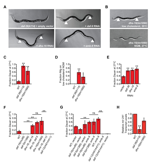
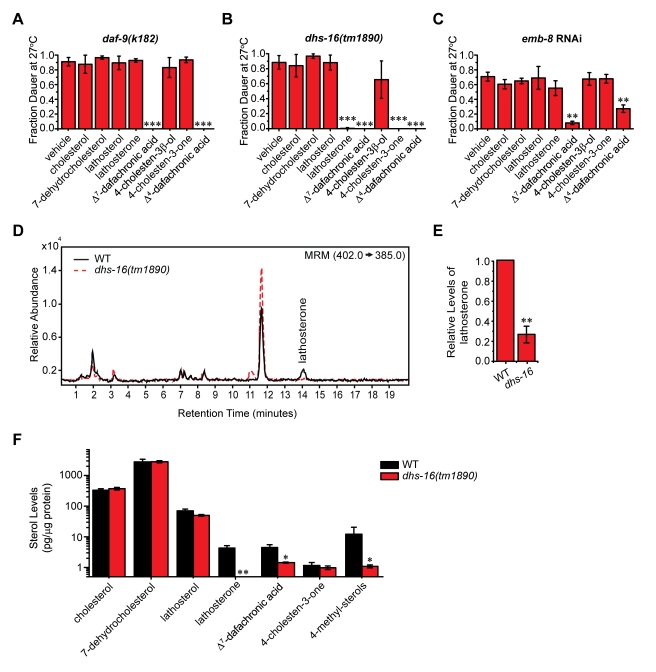
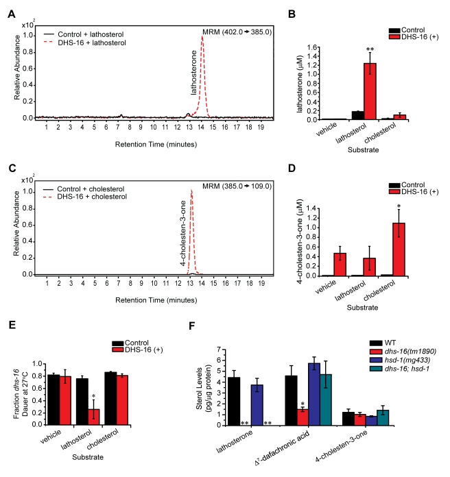
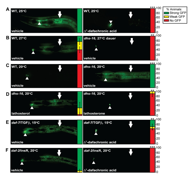
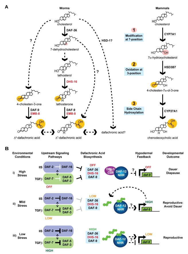
Endogenous small molecule metabolites that regulate animal longevity are emerging as a novel means to influence health and life span. In C. elegans, bile acid-like steroids called the dafachronic acids (DAs) regulate developmental timing and longevity through the conserved nuclear hormone receptor DAF-12, a homolog of mammalian sterol-regulated receptors LXR and FXR. Using metabolic genetics, mass spectrometry, and biochemical approaches, we identify new activities in DA biosynthesis and characterize an evolutionarily conserved short chain dehydrogenase, DHS-16, as a novel 3-hydroxysteroid dehydrogenase. Through regulation of DA production, DHS-16 controls DAF-12 activity governing longevity in response to signals from the gonad. Our elucidation of C. elegans bile acid biosynthetic pathways reveals the possibility of novel ligands as well as striking biochemical conservation to other animals, which could illuminate new targets for manipulating longevity in metazoans.


Conflict of interest statement

The authors have declared that no competing interests exist.

Figures

Figure 1
Figure 1. Newly identified loci display phenotypes resembling DA deficiency.
(A) Enhancement of daf-36(k114) gonadal Mig defects is seen upon knockdown of daf-9, dhs-16, and emb-8. Arrowheads indicate the arms of the gonad, visible along the body due to failure of proper distal tip cell migration. (B) Mig and Daf-c phenotypes of dhs-16(tm1890) deletion mutants, under conditions of cholesterol deprivation (upper image) and 27°C (lower image). (C) Analysis of dauer formation at 27°C on NGM. dhs-16(tm1890) mutants show Daf-c phenotypes similar to daf-36(k114) (N = 3, M±SD; **p<0.01). (D) Analysis of the gonadal Mig defects of dhs-16 null animals on NGM without added cholesterol (N = 3, M ± SD; **p<0.01, *p<0.05). (E) RNAi knockdown of dhs-16 and emb-8 in wild-type worms at 27°C induces Daf-c phenotypes, similar to knockdown of daf-36 (N≥4, M ± SD; **p<0.005). (F) Genetic epistasis analysis of dhs-16(tm1890) (Daf-c) together with Daf-d mutations in transcription factors of insulin/IGF, TGF-β, and DA signaling show that DHS-16 works downstream of DAF-16/FOXO and DAF-5/SKI, but upstream of DAF-12/NHR (N = 3, M ± SD; **p<0.01). (G) Similar genetic epistasis analysis of emb-8 RNAi-induced dauer phenotypes at 27°C suggests that EMB-8 acts downstream of DAF-16 and upstream of DAF-12 (N = 3, M ± SD; **p<0.01). (H) The DAF-12 target gene mir-241, a let-7 related microRNA, shows reduced expression in dhs-16 mutants under low cholesterol conditions at 25°C (N = 3, M ± SD; **p<0.01, *p<0.05).
Figure 2
Figure 2. dhs-16 mutant animals are deficient in lathosterone.
(A) daf-9(k182) rescue is seen only with the DAs. Experiments were carried out at 27°C with 33 µM concentration of supplemented compounds (N≥3, M ± SD; ***p<0.0001). (B) dhs-16(tm1890) rescue is seen with lathosterone and dafachronic acids, but not by cholesterol, 7-dehydrocholesterol, and lathosterol. Similarly, 4-cholesten-3-one rescues, but not cholesterol or 4-cholesten-3β-ol. This indicates that dhs-16 may function in the conversion of lathosterol to lathosterone in the production of Δ7-DA and in the formation of 4-cholestene-3-one in the production of Δ4-DA (N≥3, M ± SD; ***p<0.0001). (C) emb-8 RNAi rescue is seen only with the DAs, and not with lathosterone or 4-cholesten-3-one. (N≥3, M ± SD; **p<0.01). (D) LC/MS/MS analysis of lipid extracts from L3 stage animals reveals that lathosterone levels are reduced in dhs-16(tm1890) mutant animals compared to N2 wild-type (WT) animals, shown quantitatively in (E). Lathosterone levels are significantly reduced in dhs-16 animals relative to N2 wild-type (3.5-fold decrease; N = 7, M ± SD; **p<0.001). (F) GC/MS/MS analysis of sterol levels in dhs-16 mutants reveals deficiencies in lathosterone, Δ7-dafachronic acid, and 4-methyl sterols compared to N2 wild-type (N≥10, M ± SEM; *p<0.05, **below detection limit).
Figure 3
Figure 3. DHS-16 acts as a 3-hydroxysteroid dehydrogenase in DA biosynthesis.
(A) LC/MS/MS analysis of lipid extracts from DHS-16 and control microsomes incubated with the proposed substrate lathosterol, shown quantitatively in (B). Significantly more lathosterone is detected in incubations of DHS-16 microsomes with lathosterol than in incubations with empty vehicle ethanol controls. DHS-16 microsomes also do not produce lathosterone when incubated with cholesterol, demonstrating that specific products are made depending upon the substrate provided (N≥3, M ± SD; **p<0.005). (C) LC/MS/MS analysis of 4-cholesten-3-one levels in lipid extracts from DHS-16 and control microsomes incubated with cholesterol, shown quantitatively in (D). Concentrations of 4-cholesten-3-one in incubations of DHS-16 microsomes with cholesterol are significantly greater than that seen in control microsomes or incubations with lathosterol (N = 3, M ± SD; *p<0.05). (E) Rescue of the Daf-c phenotype of dhs-16(tm1890) mutants at 27°C is seen when fed lipid extracts of DHS-16 microsomes incubated with the proposed substrate lathosterol. Rescue is not seen with extracts from DHS-16 microsomes incubated with ethanol vehicle alone or with cholesterol, and extracts from empty vector pCMV control microsomes do not rescue in any condition (estimated concentrations of 300 nM in plates; N = 3, M ± SEM; *p<0.05). (F) GC/MS/MS analysis of sterol levels in N2 wild-type, dhs-16(tm1890), hsd-1(mg433), and dhs-16;hsd-1 double mutants reveals that hsd-1 is not required for 4-cholesten-3-one production, as previously proposed, suggesting HSD-1 may act in an alternative parallel pathway. In addition, although the dhs-16;hsd-1 double mutants did not contain measurable lathosterone levels, Δ7-DA levels were not reduced, suggesting that lathosterone is not required for its production and that alternate pathways must exist that are independent of dhs-16 (N≥6, M ± SEM; **below detection limit, *p<0.05).
Figure 4
Figure 4. dhs-16 modulates feedback regulation of hypodermal daf-9 expression.
(A) In response to mild stress (e.g., growth at 25°C), hypodermal daf-9 is upregulated in N2 wild-type animals (WT) carrying an integrated dhIs64(daf-9::gfp) array (shown at left). This is rescued upon growth with 33 µM Δ7-DA (shown at right). Arrowheads indicate the XXX R/L neuroendocrine cells in which daf-9 expression is relatively unchanged, and arrows indicate hypodermal expression. The expression levels are displayed quantitatively to the right of each image, as the percentage of animals observed with strong (green), weak (yellow), or no (red) hypodermal GFP expression (N≥3, M ± SD; ***p<0.0001). (B) Under stressful growth conditions at 27°C N2 wild-type animals still undergo reproductive development but have high hypodermal daf-9::gfp expression (left), whereas dhs-16 mutants mostly develop as dauer larvae and shut off hypodermal daf-9::gfp expression (right) (**p<0.01). (C) Under normal growth conditions at 20°C, hypodermal daf-9 upregulation is low in wild-type (left), whereas upregulation is seen in the dhs-16(tm1890) mutant background (right), suggesting daf-9 upregulation in response to DA deficiency (***p<0.0001). (D) Upregulation of hypodermal daf-9::gfp seen in dhs-16 mutants is not rescued by provision of 33 µM of the upstream DA precursor lathosterol (left) but is rescued by the downstream product lathosterone (right) (**p<0.01). (E) Upregulation of hypodermal daf-9::gfp seen in reproductively growing daf-7(e1372)/TGF-β mutants (left) is rescued by DA (right) (**p<0.01). (F) Upregulation of hypodermal daf-9::gfp seen in reproductively growing daf-2/InsR mutants (left) is also rescued by DA (right) (***p<0.0001).
Figure 5
Figure 5. dhs-16 expression pattern and regulation.
(A) A functional dhs-16::gfp is expressed in the hypodermis (arrowheads), head neurons (arrows), and posterior pharyngeal bulb (arrowheads). (B) Reduction of IIS in the daf-2(e1368) background results in 2-fold upregulation of dhs-16::gfp in the hypodermis of L3 stage animals at 20°C (*p<0.05), although no significant change was seen in the absence of daf-16/FOXO, daf-12/NHR, or daf-7/TGFβ. Similar upregulation was seen in the daf-36(k114)/Rieske oxygenase mutant background, consistent with a role of dhs-16 downstream of daf-36 in DA production (N≥3, M ± SEM, *p<0.05).
Figure 6
Figure 6. dhs-16 is partially required for longevity in the absence of the germline.
(A) Lifespan of dhs-16(tm1890) animals after ablation of germline precursor cells by laser microsurgery. One representative experiment is shown. N2 wild-type (WT) animals live twice as long when the germline is ablated. Longevity is significantly attenuated in dhs-16 ablated animals (N = 2, p<0.0001). (B) DAF-16::GFP strongly localizes to intestinal nuclei of day 1 adults after ablation of the germline by laser microsurgery in control animals, whereas the degree of localization is reduced in germline-ablated dhs-16 mutants. Magnified views of boxed regions are shown to the right. (C) The degree of DAF-16::GFP nuclear localization after germline ablation is shown quantitatively as the percentage of animals with strong (green) or weak (yellow) localization (N = 3, M ± SD; *p<0.05). (D) The ratio of intestinal DAF-16::GFP intensity in the nucleus versus cytoplasm reveals increased levels of nuclear expression in germline deficient glp-1(e2141ts) mutants at the restrictive temperature of 25°C compared to control animals. Localization is significantly decreased in dhs-16;glp-1 double mutant animals, and is restored upon provision of lathosterone or Δ7-dafachronic acid, but not lathosterol (N = 3, M ± SD; ***p<0.0001, **p<0.001).
Figure 7
Figure 7. Biosynthesis and regulation of nematode bile acids.
(A) A revised model of the dafachronic acid biosynthetic pathway from dietary cholesterol, with newly identified activities shown in red. Although dhs-16 is required for lathosterone production, mutant animals still produce low levels of Δ7-DA. An alternative pathway for Δ7-DA synthesis is therefore likely. In addition, hsd-1 is not required for 4-cholesten-3-one production as previously proposed, but may be involved in producing alternative dafachronic acids. These ligands may have complex regulation and influence the synthesis of one another. Comparison to mammalian bile acid synthesis (right) reveals conserved aspects of bile acid biochemistry. Nematode and mammalian bile acid synthesis involves modification at the 7-position (shown in pink), which speculatively may partition cholesterol towards bile acid synthesis, and in both pathways oxidation of the 3-alchohol and oxidation of the sidechain at the 27-position occurs (shown in orange). (B) Model of hormonal feedback on hypodermal daf-9::gfp expression. (i) Stressful environmental conditions result in downregulation of IIS and TGF-β signaling, suppression of DA synthesis, and hypodermal daf-9 expression by the DAF-12/DIN-1 repressor complex, resulting in dauer formation. (ii) Moderately stressful environments result in modest downregulation of dauer signaling pathways and DA synthesis, with compensatory upregulation of hypodermal daf-9, allowing for reproductive development. (iii) In favorable environments with low levels of stress, active IIS and TGF-β signaling results in ample DA production, with low expression of hypodermal daf-9. Note it is unknown whether DAF-12 regulates daf-9 directly or indirectly.

Similar articles

Cited by

References

    1. Kenyon C. J. The genetics of ageing. Nature. 2010;464:504–512. - PubMed
    1. Eisenberg T, Knauer H, Schauer A, Buttner S, Ruckenstuhl C, et al. Induction of autophagy by spermidine promotes longevity. Nat Cell Biol. 2009;11:1305–1314. - PubMed
    1. Honda Y, Tanaka M, Honda S. Trehalose extends longevity in the nematode Caenorhabditis elegans. Aging Cell. 2010;9:558–569. - PubMed
    1. Lucanic M, Held J. M, Vantipalli M. C, Klang I. M, Graham J. B, et al. N-acylethanolamine signalling mediates the effect of diet on lifespan in Caenorhabditis elegans. Nature. 2011;473:226–229. - PMC - PubMed
    1. Goudeau J, Bellemin S, Toselli-Mollereau E, Shamalnasab M, Chen Y, et al. Fatty Acid Desaturation Links Germ Cell Loss to Longevity Through NHR-80/HNF4 in C. elegans. PLoS Biol. 2011;9:e1000599. - PMC - PubMed

Publication types

MeSH terms