. 2012:2012:409496.
doi: 10.1155/2012/409496. Epub 2012 Feb 6.

Application of laser-induced bone therapy by carbon dioxide laser irradiation in implant therapy

Affiliations

Application of laser-induced bone therapy by carbon dioxide laser irradiation in implant therapy

Takahiro Naka et al. Int J Dent. 2012.

Abstract

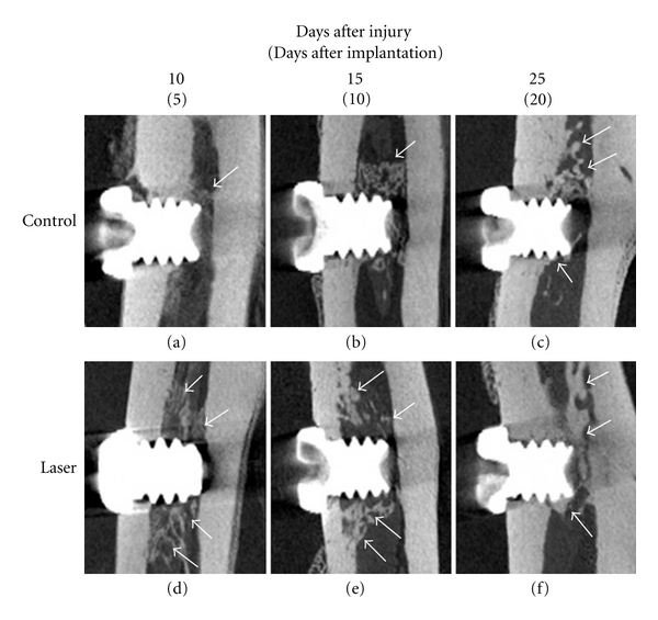
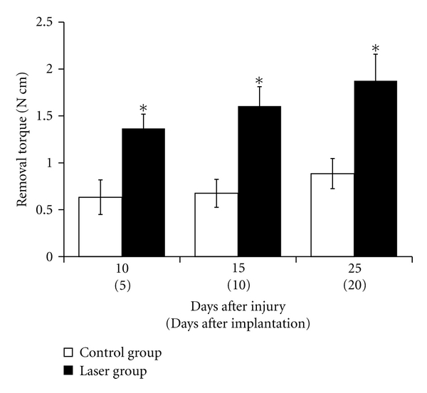
This study evaluated the application of laser-induced bone therapy (LIBT) to reduce implant healing time in rat tibia. Twenty 10-week-old female Sprague-Dawlay rats were used. The rats received laser irradiation (laser group) or sham operation (control group) on either side of the tibia. Five days after invasion, titanium implants were inserted in proximal tibia. Five, 10, and 20 days after implant placement, tibiae were collected. After taking micro-CT and performing a torque test, the tibiae were decalcified and 8-μm-thick sections were prepared. Specimens were stained with hematoxylin and eosin. Results. Micro-CT images, removal torque values, and histomorphometric analysis data demonstrated a significantly accelerated bone formation in the laser group earlier in the healing process. Conclusion. The use of laser irradiation was effective in promoting bone formation and acquiring osseointegration of titanium implants inserted in rat tibia. LIBT may be suitable for use in implant therapy.


Figures

Figure 1
Figure 1
Experimental protocol. Time schedule of bur/laser injury, implant placement, and subsequent healing periods.
Figure 2
Figure 2
Comparison of osteoid formation in the bone marrow space in the laser-irradiated group and bur-injured group 5 days after treatment. (a) laser-irradiated tibia. Energy density = 88.2 J/cm2, (output: 1.0 W, irradiation time: 2 sec). (b) laser-irradiated tibia. Energy densities = 220.5 J/cm2, (output: 1.0 W, irradiation time: 5 sec). (c) laser-irradiated tibia. Energy densities = 441.0 J/cm2, (output: 1.0 W, irradiation time: 10 sec). (d) laser-irradiated tibia. Energy densities = 661.5 J/cm2, (output: 1.0 W, irradiation time: 15 sec). (e) bur-injured tibia. (a–d) laser-irradiated tibia showed an ablation defect, carbon deposits, and numerous empty osteocytic lacunae. Moreover, newly formed trabecular bone was observed on the marrow side of the laser-treated site (a–c). (e) bur-injured tibia showed a slight amount of reactive bone formation on the endosteal surface. H&E stain with 40x magnification. Bar = 200 μm. Arrow indicates laser-irradiated or bur-injured site. Arrow heads indicate osteoid formation site.
Figure 3
Figure 3
Micro-CT images. (a, d): 5 days after implantation (10 days after sham operation or laser irradiation). (b, e): 10 days after implantation (15 days after sham operation or laser irradiation). (c, f): 20 days after implantation (25 days after sham operation or laser irradiation). (a–c): control group tibia. (d–f): laser group tibia. Arrow indicates newly formed bone.
Figure 4
Figure 4
Removal torque examination of titanium implant. Removal torque values were measured at different time points. Mean ± SE (N cm) of torque forces in laser group and control group. Five days after implantation, significant difference was observed between the control and laser groups. Until twenty days after implantation, there was still a significant difference between the two groups. *P < 0.05.
Figure 5
Figure 5
H&E staining observation. (a, d): 5 days after implantation (10 days after sham operation or laser irradiation). (b, e): 10 days after implantation (15 days after sham operation or laser irradiation). (c, f): 20 days after implantation (25 days after sham operation or laser irradiation). (a–c): control group tibia. (d–f): laser group tibia. In the early healing period (10 days after injury), in the control group, there was limited bone formation around the implant body, while in the laser group, there was obvious bone formation along the inserted implant. Fifteen days after injury, in the control group, tibia showed little osteoid formation around the implant body. In the laser group, thick and abundant bone formation was evident along the inserted implant body. Twenty-five days after injury, in the control group, tibia showed newly formed cortical bone, but the trabecular bone was thinner than that in the laser group tibia. In the laser group, thick cortical bone was connected to the previously existing cortex. H&E stain with 40x magnification. c: cortical bone, i: implant cavity, nb: newly formed bone, bar = 200 μm.
Figure 6
Figure 6
Summary of the healing processes in the control group and laser group. Laser irradiation-induced bone formation in the bone marrow and laser group showed earlier osseointegration. The control group also acquired osseointegration, but the healing period was longer than that of the laser group.

References

    1. Brånemark PI, Zarb GA, Albrektsson T. Tissue Integrated Protheses: Osseointegration in Clinical Dentistry. Chicago, Ill, USA: Quintessence; 1985.
    1. Adell R, Lekholm U, Rockler B, Branemark PI. A 15-year study of osseointegrated implants in the treatment of the edentulous jaw. International Journal of Oral Surgery. 1981;10(6):387–416. - PubMed
    1. Albrektsson T, Zarb G, Worthington P, Eriksson AR. The long-term efficacy of currently used dental implants: a review and proposed criteria of success. The International Journal of Oral &amp; Maxillofacial Implants. 1986;1(1):11–25. - PubMed
    1. Wolff J. Das Gesetz der Transformation der Knochen. Berlin, Germany: Springer; 1892. Translated as The Law of Bone Remodeling.
    1. Frost HM. Wolff’s Law and bone’s structural adaptations to mechanical usage: an overview for clinicians. Angle Orthodontist. 1994;64(3):175–188. - PubMed

LinkOut - more resources