. 2012 Mar;6(1):14116-1411611.
doi: 10.1063/1.3692765. Epub 2012 Mar 7.

Microfluidic sorting of microtissues

Microfluidic sorting of microtissues

D G Buschke et al. Biomicrofluidics. 2012 Mar.

Abstract

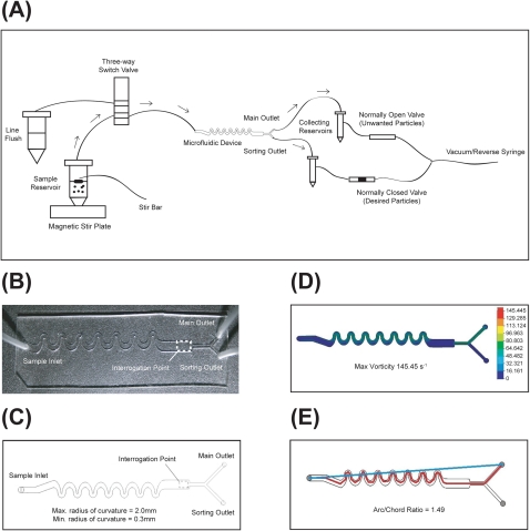
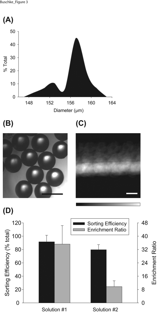
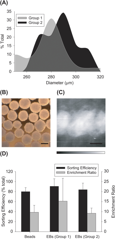
Increasingly, invitro culture of adherent cell types utilizes three-dimensional (3D) scaffolds or aggregate culture strategies to mimic tissue-like, microenvironmental conditions. In parallel, new flow cytometry-based technologies are emerging to accurately analyze the composition and function of these microtissues (i.e., large particles) in a non-invasive and high-throughput way. Lacking, however, is an accessible platform that can be used to effectively sort or purify large particles based on analysis parameters. Here we describe a microfluidic-based, electromechanical approach to sort large particles. Specifically, sheath-less asymmetric curving channels were employed to separate and hydrodynamically focus particles to be analyzed and subsequently sorted. This design was developed and characterized based on wall shear stress, tortuosity of the flow path, vorticity of the fluid in the channel, sorting efficiency and enrichment ratio. The large particle sorting device was capable of purifying fluorescently labelled embryoid bodies (EBs) from unlabelled EBs with an efficiency of 87.3% ± 13.5%, and enrichment ratio of 12.2 ± 8.4 (n = 8), while preserving cell viability, differentiation potential, and long-term function.


Figures

Figure 1
Figure 1
Characterization of the LaPSD. (a) Schematic depicting the particle flow path. Black arrows denote particle path during main flow. Dotted arrows denote particle path during a sorting event. (b) Top view of the LaPSD design consisting of one inlet port, an interrogation region, a main outlet port, and a sorting outlet port. (c) Photograph depicting the fabricated LaPSD device, formed by bonding PDMS to glass cover slip. (d) 2D colorimetric vorticity plot of the LaPSD. The maximum vorticity during main flow (analogous to sorting flow) is 145 s−1. (e) Tortuosity of the LaPSD as defined by the arc to chord ratio is 1.49.
Figure 2
Figure 2
Viability and long-term function of stem cell aggregates following flow through the LaPSD. Fluorescently labelled EBs were introduced into the device through the sample inlet, optically interrogated and removed via the main outlet. After removal, EBs were placed in culture plates and the number of EBs that attached were counted and reported as a percentage of the total number of EBs plated compared to control EBs that were generated at the same time, but placed immediately in static culture dishes. In addition, EBs were tracked over time and the percentage of EBs with functional cardiomyocytes (i.e., beating areas) was determined. Attachment and beating functionality were not significantly altered with flow through the LaPSD compared to controls (P = 0.60 and 0.33, respectively). Error bars correspond to standard deviation from the mean (enhanced online) .
Figure 3
Figure 3
Sorting efficiency and enrichment ratio of polystyrene beads. (a) Histogram depicting the distribution of bead diameters in a representative fraction of fluorescent and non-fluorescent polystyrene beads. Measured bead diameter (157 ± 3 μm) did not differ statistically from manufacturer’s specifications (158 ± 3 μm). (b) Brightfield image of polystyrene beads. Scale bar = 200 μm. (c) Summation of temporal image series of beads flowing through the LaPSD. Fluorescence intensity images of beads were taken every 0.2 s and ∼3000 images were merged to create the image shown. The minimal deviation of fluorescence intensity from the center sample stream line conveys the ability of the LaPSD to effectively focus particles with uniform size. Scale bar = 200 μm. (d) Sorting efficiency and enrichment ratio of bead populations consisting of fluorescent and nonfluorescent beads. Solution 1 attained significantly higher enrichment ratios due to a low starting concentration of fluorescent beads; this result demonstrates the ability of the LaPSD to effectively purify particles from relatively dilute starting concentrations akin to rare cell populations. Error bars correspond to standard deviation from the mean.
Figure 4
Figure 4
Sorting efficiency and enrichment ratio of embryoid bodies. (a) Histograms depicting measured EB diameters for two separate groups of EBs. Measured mean EB diameters were 247 ± 20 μm for group 1, and 295 ± 12 μm for group 2. (b) Brightfield images of EBs. Scale bar = 200 μm. (c) Summation of temporal image series of EBs flowing through the LaPSD. Fluorescence intensity images of EBs were taken every 0.2 s and ∼2000 images were merged to create the image shown. The deviation of fluorescence intensity from the center sample stream line is more diffuse than that observed for beads reflecting the higher variability of size of EBs and therefore greater perturbation of flow. Scale bar = 200 μm. (d) Sorting efficiency and enrichment ratio of EB groups compared to that of bead solution 2. There was no significant difference between sorting efficiency or enrichment ratios when comparing beads to either group of EBs. Error bars correspond to standard deviation from the mean.

Similar articles

Cited by

References

    1. Leong D. T., Nah W. K., Gupta A., Hutmacher D. W., and Woodruff M. A., Curr. Drug Discovery Technol. 5, 319 (2008).10.2174/157016308786733537 - DOI - PubMed
    1. Phillips B. W., Horne R., Lay T. S., Rust W. L., Teck T. T., and Crook J. M., J. Biotechnol. 138, 24 (2008).10.1016/j.jbiotec.2008.07.1997 - DOI - PubMed
    1. Carpenedo R. L., Bratt-Leal A. M., Marklein R. A., Seaman S. A., Bowen N. J., McDonald J. F., and McDevitt T. C., Biomaterials 30, 2507 (2009).10.1016/j.biomaterials.2009.01.007 - DOI - PMC - PubMed
    1. Stegemann J. P. and Nerem R. M., Exp. Cell Res. 283, 146 (2003).10.1016/S0014-4827(02)00041-1 - DOI - PubMed
    1. Schmeichel K. L. and Bissell M. J., J. Cell Sci. 116, 2377 (2003).10.1242/jcs.00503 - DOI - PMC - PubMed

LinkOut - more resources