. 2012;7(4):e34596.
doi: 10.1371/journal.pone.0034596. Epub 2012 Apr 10.

MicroRNAs overexpressed in growth-restricted rat skeletal muscles regulate the glucose transport in cell culture targeting central TGF-β factor SMAD4

Affiliations

MicroRNAs overexpressed in growth-restricted rat skeletal muscles regulate the glucose transport in cell culture targeting central TGF-β factor SMAD4

Santanu Raychaudhuri. PLoS One. 2012.

Abstract

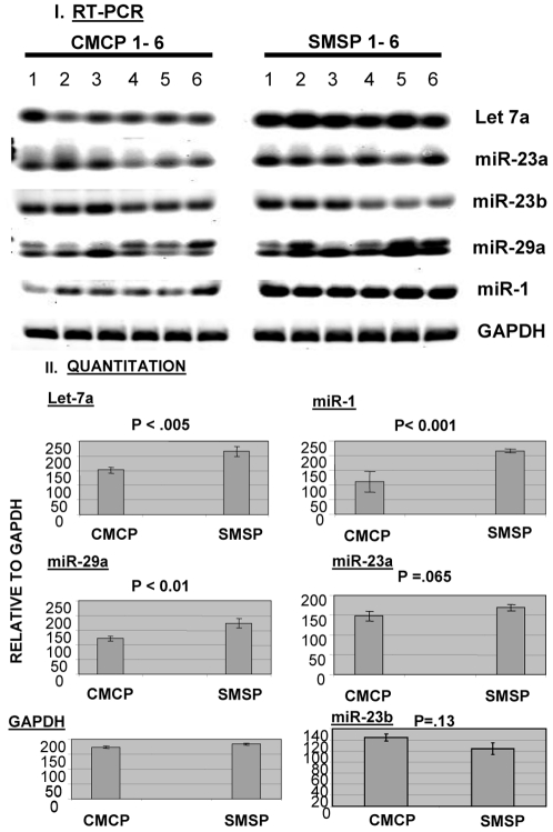
The micro-array profiling of micro-RNA has been performed in rat skeletal muscle tissues, isolated from male adult offspring of intrauterine plus postnatal growth restricted model (IPGR). Apparently, the GLUT4 mRNA expression in male sk. muscle was found to be unaltered in contrast to females. The over-expression of miR-29a and miR-23a in the experimental group of SMSP (Starved Mother Starved Pups) have been found to regulate the glucose transport activity with respect to their control counterparts CMCP (Control Mother Control Pups) as confirmed in rat L6 myoblast-myocyte cell culture system. The ex-vivo experimentation demonstrates an aberration in insulin signaling pathway in male sk. muscle that leads to the localization of the membrane-bound Glut4 protein. We have identified through a series of experiments one important protein factor SMAD4, a co-SMAD critical to the TGF-beta signaling pathway. This factor is targeted by miR-29a, as identified in an in vitro reporter-assay system in cell-culture experiment. The other micro-RNA, miR-23a, targets SMAD4 indirectly that seems to be critical in regulating insulin-dependent glucose transport activity. MicroRNA mimics, inhibitors and siRNA studies indicate the role of SMAD4 as inhibitory for glucose transport activities in normal physiological condition. The data demonstrate for the first time a critical function of microRNAs in fine-tuning the regulation of glucose transport in skeletal muscle. Chronic starved conditions (IPGR) in sk. muscle up-regulates microRNA changing the target protein expression patterns, such as SMAD4, to alter the glucose transport pathways for the survival. The innovative outcome of this paper identifies a critical pathway (TGF-beta) that may act negatively for the mammalian glucose transport machinery.


Conflict of interest statement

Competing Interests: The author has declared that no competing interests exist.

Figures

Figure 1
Figure 1. Glut4 mRNA expression level does not alter with the IPGR semi-calorie food restricted male rat skeletal muscles.
A. Representative Northern blot analysis using total RNA isolated from six male rat SKM samples (450 days-IPGR) of both control (CMCP) and food restricted animals (SMSP). GLUT4 and β-Actin specific probes were used in hybridization. Ethidium bromide stained 28S and 18S ribosomal RNA is used as loading control. β-Actin is used as internal control. B. Intensity of the band was quantitated and expressed as percentages. C. Real-time PCR has been performed using the same RNA samples (1–6) of CMCP and SMSP male rat SKM using GLUT4 specific TaqMan probe and primers using GAPDH, Glyceraldehyde-3-phosphate dehydrogenase, as internal control and D. Average of six samples.
Figure 2
Figure 2. Over-expression of some microRNAs is observed in IPGR SKM samples over the control animals.
Differential expression profile of sixty micro-RNAs that bind to the Chips in LNA-based microarray analysis (Exiqon miRNA-vol 8.1) of male rat SKM micro-RNA enriched samples isolated from CMCP (CC1-6) and SMSP (SS1-6) animals. CC1-6 samples are labeled with Hy3 and SS1-6 by Hy5 dyes. Log2ratio of Hy3 by Hy5 is plotted against every bound microRNAs. Sample size was N = 3 and 10 different spike-in controls have been used for the experiment as internal control.
Figure 3
Figure 3. Semi-quantitative RT-PCR analysis of microRNAs in male SKM samples.
I. The total RNA (1 µg) has been used to make cDNA using Ambion mirVana isolation kit. Equal amount of cDNA has been used in separate microRNA-specific PCR reactions. CMCP (1-6) and SMSP (1-6) correspond to six samples of control and starved SKM. II. Image J analyses of the intensities of all bands were plotted as graphs for each microRNAs. T-test was done as a measure of statistical analysis. GAPDH was used as an internal control.
Figure 4
Figure 4. Analyses of mature microRNAs of interest found in both micro-array and RT-PCR analysis.
Denaturing Urea-Acrylamide gel electrophoresis of total RNA (10 µg) have been run and subjected to Northern Blot analysis using P32-labelled LNA-based microRNA probes (Exiqon) either as mixed samples of 6 SKM RNA (I) or separately for 6 samples for miR-1 (II). The single arrow stands for the mature microRNA of appropriate size and the double arrow signifies the precursor forms, 60–100 bases. SMSP is the experimental against CMCP as control animal samples. III. Relative intensities of the bands in I and II.
Figure 5
Figure 5. Cellular fractionation of possible target proteins based on microRNA bioinformatics analysis and the insulin-signaling proteins for glucose transport: Membrane fraction.
The biochemical fractionations of the membrane proteins were made using Chemicon/Millipore's compartmental sub-cellular analysis technology. Cytosolic, membrane, nuclear and cytoskeleton fractions have been made from SKM tissues from both control (CMCP) and experimental (SMSP) male animals. SDS-PAGE followed by Western Blot analysis was made using 50 µg of total protein from every fractions based on Bradford assay. The localization of GLUT4 and IGFR in IPGR male SKM is significantly increased in membrane fractions when the total GLUT4 protein level remains unaltered in male SKM. GLUT1, 3 and 4 are Slc2 group of glucose transport proteins, IGFR is Insulin Growth Factor Receptor protein, EGFR is used as internal control for membrane localized protein. Vamp2 and Vimentin are two essential regulatory protein markers for insulin signaling and cell differentiation. β-Actin was used as internal control for total protein. The bottom panel represents the verification data for the cell fractionation. GAPDH, Nucleolin and EGFR were used for cytosolic (C), nuclear (N) and membrane (M) localized proteins. T represents total protein.
Figure 6
Figure 6. Cellular fractionation of possible target proteins: Cytosolic fraction.
Insulin signaling marker is up-regulated along with the metabolic marker glucose-6-phospahe dehydrogenase and ARF1 GTPase, whereas SKM marker Tropomyocin C is down-regulated along with two essential signaling candidates SMAD4 and DnaJ-B1.
Figure 7
Figure 7. Cellular fractionation of possible target proteins: Cytoskeletal fraction.
Insulin-signaling protein VAMP2 is upregulated along with the regulatory protein Vimentin, whereas SMAD4 TGF-β co-SMAD is down-regulated. NSP represents non-specific protein serving as internal control.
Figure 8
Figure 8. SMAD4, is one of the targets for miR-29a and/or miR-23a: Bioinformatics analysis.
miR-1, miR-29a and miR-23a for SMAD4, DnaJ-B1 and nucleolin 3′UTRs. MIRANDA and Sloan-Kettering MicroRNA target analysis site were used for this analysis. Alignment score, PhastCons score and Energy values were cited for all the interactions.
Figure 9
Figure 9. Mimic microRNAs miR-29a and miR-23 up-regulate the glucose transport activities in L6 SKM cell-line as insulin-independent and dependent manner respectively.
A. Pre-miR transient transfection. 25 nM Pre-miRs were transfected in rat L6 cells according to the protocol as described in method section. 14C-labelled 2-deoxyglucose and 3H-labelled OMe-glucose were used as substrate along with the non-radioactive substrate (0.15 mM) for glucose transport activity as reversible and irreversible manner respectively. Cytochalasin B is not being added. B. Pre-miR transfection with Insulin. Same transfection experiments were done with cytochalasin B (10 µM) added in the transport reaction in presence and absence of 50 nM insulin. Glucose transport assay was performed just (I) before and (II) after adding insulin. (III) Panel was drawn to see the comparison before and after the insulin addition for each mimic precursor microRNA with respect to the control transport of 14C-labelled 2-deoxyglucose.
Figure 10
Figure 10. MicroRNA anti-sense inhibitors (miR-29a and miR-23a) down-regulate the glucose transport activity in insulin-added condition in L6 cell-line.
Ambion-tested specific endogenous microRNA inhibitors (25 nM) were used in this experiment basically the same way as mimic transfection experiment (A & B). Only, the standard radioactivity amount was increased to 7.5 µCi per 12 well plate for both (I) without insulin and (II) with insulin conditions. Cytochalasin B (10 µM) was added in the transport reactions.
Figure 11
Figure 11. Cellular fractionation of possible target proteins: Nuclear fraction.
Nucleolin, a nuclear regulatory factor and HDAC4 and MEF2C, the transcription factors, are down-regulated, whereas HAND2 and SMAD4 remain unaltered. Lamin A served as internal control.
Figure 12
Figure 12. SMAD4 is one of the targets for miR-29a and/or miR-23a: Analysis in Cell-line.
PremiR transfections (25 nM) of rat L6 myoblast-myocytes were done and the cell-extracts were subjected to Western Blot analysis for SMAD4, nucleolin and DnaJ-B1 level after 48 hours (I). The triplicate experiments were run in SDS-PAGE/Western Blot to have statistical significant data (T-test) and expressed as relative intensities (%) (I).
Figure 13
Figure 13. Reporter analysis for microRNA targeting SMAD4 3′UTR.
The 3′UTR was cloned in luciferase based pMIR-reporter of Ambion for reporter analysis of microRNA binding sites in its UTR. Plasmid was transfected in presence and absence of precursor microRNAs; 29a, 1 and 23a mimic (25 nM). (I) 3 clones were transfected separately w.r.t. vector without SMAD4 3′UTR. (II) Pre-miR-29a, (III) Pre-miR-23a and (IV) Pre-miR-1 precursors were transfected along with either SMAD4-3′UTR clone or empty vector. Luciferase light units (RLU) were measured in luminometer and expressed as per OD(595) protein measured by Bradford. Triplicate was assayed and expressed as statistical significance by T-test.
Figure 14
Figure 14. Depletion of SMAD4 activates the glucose transport activity in both skeletal muscle and cardiomyocyte cells.
Small inhibitory RNA (25 nM) was used to deplete SMAD4 in L6 cell culture system prior to glucose transport assay using 14C-2-deoxyglucose as described in method. Both skeletal muscle cells (I & II) and cardiomyocytes cells (III) were used for this experiment. The transport assay condition is maintained in the same way as was done for Fig. 3. (IV) The Western Blot analysis in L6 cell-lines to show the specificity of siRNA against SMAD4. Dharmacon-designed SMARTpool siRNAs (4 sets) specific to SMAD4 and non-targeting negative control siRNA were used for this experiment according to the manufacturer's instruction.

References

    1. Callis TE, Chen JF, Wang DZ. DNA Cell Biology. DNA Cell Biology. 2007;26(4):219–25. - PubMed
    1. Williams AH, Liu N, van Rooij E, Olson EN. MicroRNA control of muscle development and disease. Curr Opin Cell Biol. 2009;21(3):461–9. - PMC - PubMed
    1. Townley-Tilson WH, Callis TE, Wang D. MicroRNAs 1, 133, and 206: critical factors of skeletal and cardiac muscle development, function, and disease. Int J Biochem Cell Biol. 2010;42(8):1252–5. - PMC - PubMed
    1. Hoffman NJ, Elmendorf JS. Signaling, cytoskeletal and membrane mechanisms regulating GLUT4 exocytosis. Trends Endocrinol Metab. 2011;22(3):110–6. - PMC - PubMed
    1. Leney SE, Tavaré JM. The molecular basis of insulin-stimulated glucose uptake: signaling, trafficking and potential drug targets. J Endocrinol. 2009;203(1):1–18. - PubMed

Publication types

MeSH terms