

Cardiomyocyte microvesicles contain DNA/RNA and convey biological messages to target cells
- PMID: 22506041
- PMCID: PMC3323564
- DOI: 10.1371/journal.pone.0034653
Cardiomyocyte microvesicles contain DNA/RNA and convey biological messages to target cells
Abstract
Background: Shedding microvesicles are membrane released vesicles derived directly from the plasma membrane. Exosomes are released membrane vesicles of late endosomal origin that share structural and biochemical characteristics with prostasomes. Microvesicles/exosomes can mediate messages between cells and affect various cell-related processes in their target cells. We describe newly detected microvesicles/exosomes from cardiomyocytes and depict some of their biological functions.
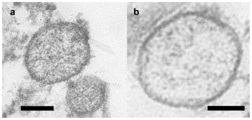
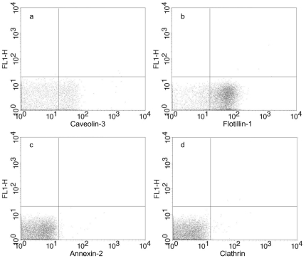
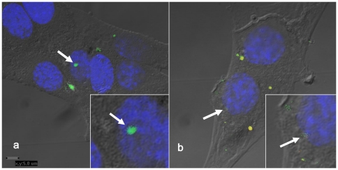
Methodology/principal findings: Microvesicles/exosomes from media of cultured cardiomyocytes derived from adult mouse heart were isolated by differential centrifugation including preparative ultracentrifugation and identified by transmission electron microscopy and flow cytometry. They were surrounded by a bilayered membrane and flow cytometry revealed presence of both caveolin-3 and flotillin-1 while clathrin and annexin-2 were not detected. Microvesicle/exosome mRNA was identified and out of 1520 detected mRNA, 423 could be directly connected in a biological network. Furthermore, by a specific technique involving TDT polymerase, 343 different chromosomal DNA sequences were identified in the microvesicles/exosomes. Microvesicle/exosomal DNA transfer was possible into target fibroblasts, where exosomes stained for DNA were seen in the fibroblast cytosol and even in the nuclei. The gene expression was affected in fibroblasts transfected by microvesicles/exosomes and among 333 gene expression changes there were 175 upregulations and 158 downregulations compared with controls.
Conclusions/significance: Our study suggests that microvesicles/exosomes released from cardiomyocytes, where we propose that exosomes derived from cardiomyocytes could be denoted "cardiosomes", can be involved in a metabolic course of events in target cells by facilitating an array of metabolism-related processes including gene expression changes.
Conflict of interest statement
Figures
References
-
- Ronquist G, Brody I, Gottfries A, Stegmayr B. An Mg2+ and Ca2+-stimulated adenosine triphosphatase in human prostatic fluid–part II. Andrologia. 1978;10:427–433. - PubMed
-
- Ronquist G, Brody I, Gottfries A, Stegmayr B. An Mg2+ and Ca2+-stimulated adenosine triphosphatase in human prostatic fluid: part I. Andrologia. 1978;10:261–272. - PubMed
-
- Ronquist G, Hedstrom M. Restoration of detergent-inactivated adenosine triphosphatase activity of human prostatic fluid with concanavalin A. Biochim Biophys Acta. 1977;483:483–486. - PubMed
-
- Ronquist G, Brody I. The prostasome: its secretion and function in man. Biochim Biophys Acta. 1985;822:203–218. - PubMed
-
- Sahlen GE, Egevad L, Ahlander A, Norlen BJ, Ronquist G, et al. Ultrastructure of the secretion of prostasomes from benign and malignant epithelial cells in the prostate. Prostate. 2002;53:192–199. - PubMed
Publication types
MeSH terms
Substances
LinkOut - more resources
Full Text Sources
Other Literature Sources
