. 2012;7(4):e35005.
doi: 10.1371/journal.pone.0035005. Epub 2012 Apr 10.

The immune response to melanoma is limited by thymic selection of self-antigens

Affiliations

The immune response to melanoma is limited by thymic selection of self-antigens

Ulrike Träger et al. PLoS One. 2012.

Abstract

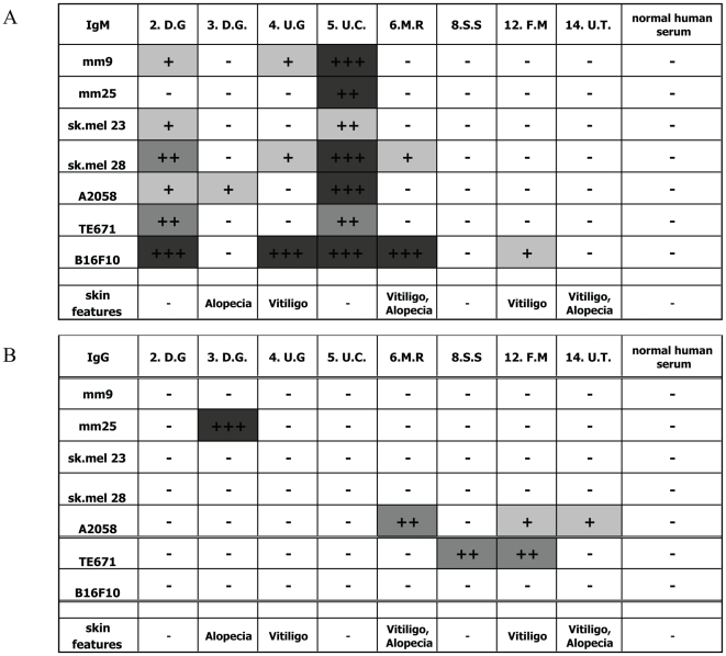
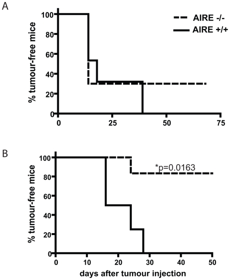
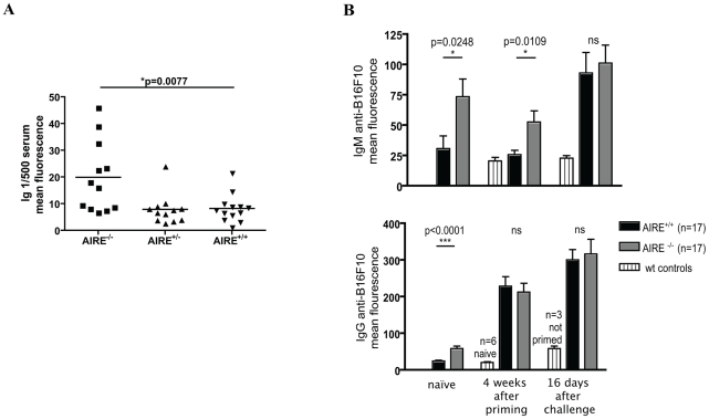
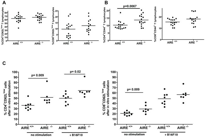
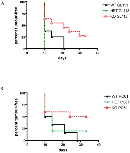
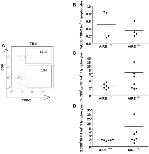
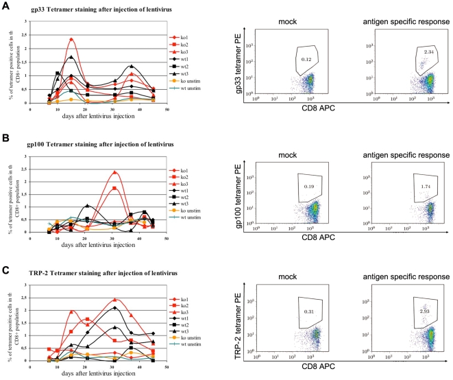
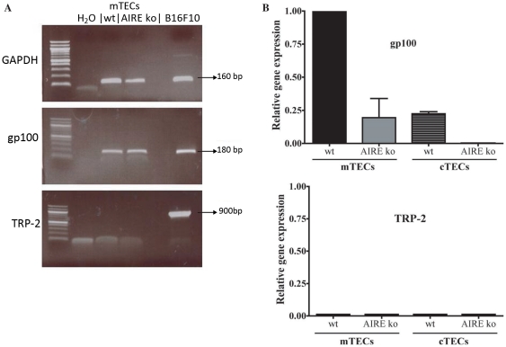
The expression of melanoma-associated antigens (MAA) being limited to normal melanocytes and melanomas, MAAs are ideal targets for immunotherapy and melanoma vaccines. As MAAs are derived from self, immune responses to these may be limited by thymic tolerance. The extent to which self-tolerance prevents efficient immune responses to MAAs remains unknown. The autoimmune regulator (AIRE) controls the expression of tissue-specific self-antigens in thymic epithelial cells (TECs). The level of antigens expressed in the TECs determines the fate of auto-reactive thymocytes. Deficiency in AIRE leads in both humans (APECED patients) and mice to enlarged autoreactive immune repertoires. Here we show increased IgG levels to melanoma cells in APECED patients correlating with autoimmune skin features. Similarly, the enlarged T cell repertoire in AIRE(-/-) mice enables them to mount anti-MAA and anti-melanoma responses as shown by increased anti-melanoma antibodies, and enhanced CD4(+) and MAA-specific CD8(+) T cell responses after melanoma challenge. We show that thymic expression of gp100 is under the control of AIRE, leading to increased gp100-specific CD8(+) T cell frequencies in AIRE(-/-) mice. TRP-2 (tyrosinase-related protein), on the other hand, is absent from TECs and consequently TRP-2 specific CD8(+) T cells were found in both AIRE(-/-) and AIRE(+/+) mice. This study emphasizes the importance of investigating thymic expression of self-antigens prior to their inclusion in vaccination and immunotherapy strategies.


Conflict of interest statement

Competing Interests: The authors have declared that no competing interests exist.

Figures

Figure 1
Figure 1. APECED patients with skin features have increased anti-melanoma antibody levels.
Cell lines were stained with serum obtained from APECED patients (2.D.G, 3.D.G., 4.U.G., 5.U.C., 6.M.R., 8.S.S., 12.F.M., 14.U.T.) diluted 1/500. Mm9, mm25, sk.mel23, sk.mel28, A2058 are human melanoma lines, TE671 a human rhabdomyosarcoma, and B16F10 murine melanoma cell line. Background using normal human serum was substracted and mean fluorescence indicated as follows −<0.5, +0.5–0.75, ++0.75–1, +++>1. The secondary antibodies used were A anti-human IgM FITC and B anti-human IgG FITC.
Figure 2
Figure 2. AIRE−/− mice reject tumour more efficiently than AIRE+/+ littermate controls mice after priming.
A Mice of indicated genotype were challenged with B16F10, and tumour growth monitored over 75 days. B As treated in A, but mice were primed with irradiated B16F10 4 weeks prior to challenge. Curves were compared with Logrank test with p values indicated. Similar trends were found in at least 5 different experiments. In each of these experiments between 5 and 10 mice/group were included.
Figure 3
Figure 3. Anti-B16F10 antibodies serum levels are elevated.
Mice of the indicated genotype were primed with 5×10⁶ irradiated B16F10 and challenged with 2.5×10⁵ live B16F10. A At time of sacrifice (when tumours reached 10 mm2 or in the case of tumour-free mice after 50 days), serum was obtained. 1/500 diluted serum was used to stain B16F10. Secondary antibody was an anti-mouse pan Ig APC. B Serum was obtained before priming, 4 weeks after priming, or 16 days after live tumour challenge and used to stain B16F10. Naïve littermate controls or unprimed but challenged controls were included were indicated. Secondary antibodies used were anti-mouse IgM FITC (upper panel) or anti-mouse IgG FITC (lower panel). * p value by Mann-Whitney test comparing mean fluorescence of serum staining B16F10. These findings were reproduced three times with similar results with n>10 in each group.
Figure 4
Figure 4. AIRE−/− mice have increased overall CD4 and CD8 responses after tumour challenge.
Mice of indicated genotype were primed and challenged with B16F10, then splenocytes were isolated, and stained for CD62L or CD44. A Percentage of CD62Llow cells among CD4+ or CD8+ T lymphocytes ex vivo B Percentage of CD44+ cells among CD4+ or CD8+ T lymphocytes ex vivo C Percentage of CD62Llow cells among CD4+ or CD8+ T lymphocytes after in vitro culture with IFNγ stimulated B16F10. * p value Mann-Whitney test comparing % of cells with low CD62L among CD4+ CD3+ T lymphocytes. This result is representative of two repeats.
Figure 5
Figure 5. Depletion of regulatory T cells leads to improved tumour rejection in both AIRE−/− and AIRE+/+ mice.
Mice of the indicated genotype were primed and challenged with B16F10. Three days and one day prior to challenge, mice were treated twice with 0.5 mg of control antibody GL113 (A), or with regulatory T cell-depleting antibody PC61 (B). Seven days after depletion with PC61 but not GL113 antibodies, CD4+CD25+ cells were reduced by 60% in the blood. Appearance of tumours was measured for 121 days.
Figure 6
Figure 6. MAA-specific CD8+ T cells present in TILs and draining LNs.
A Wildtype mice were injected with 106 live B16F10 and 10 days later, TILs were isolated and stained for CD8 and TRP-2-specific TCR using tetramers. B and C Mice of indicated genotypes were primed and challenged with B16F10 cells, then frequencies of gp100, TRP-1 or TRP-2 tetramer positive cells in the CD8+ T cell population were plotted. Representative of two similarly conducted experiments.
Figure 7
Figure 7. AIRE−/− mice do not hyperreact to foreign antigen but respond better to certain melanocyte antigen than AIRE+/+ littermate controls.
Mice of the indicated genotype were injected s.c at the base of the tail with 4×10⁶ pfu of lentivirus expressing the indicated peptides. Serial samples of blood cells (left panels) were stained for CD8 and MHC class I tetramers specific for T cells responding to the injected peptides (example at peak of response in right panels). A LCMV-derived foreign gp33 presented by H-2Db B mouse TRP-2 presented by H-2Kb C gp100 presented by H-2Db.
Figure 8
Figure 8. Expression of melanocyte antigens in medullary thymic epithelial cells.
Medullary or cortical thymic epithelial cells were sorted by flow cytometry. A RT-PCR was performed on both types of cells using primers for GAPDH, gp100 and TRP-2. B Real time qPCR was performed using sorted populations. Expression relative to gp100 expression in AIRE+/+ mTECs is shown for gp100 and TRP-2. Error bars are from duplicates.

References

    1. Miller AJ, Mihm MC., Jr Melanoma. N Engl J Med. 2006;355:51–65. - PubMed
    1. Castelli C, Rivoltini L, Andreola G, Carrabba M, Renkvist N, et al. T-cell recognition of melanoma-associated antigens. J Cell Physiol. 2000;182:323–331. - PubMed
    1. Overwijk WW, Restifo NP. Autoimmunity and the immunotherapy of cancer: targeting the “self” to destroy the “other”. Crit Rev Immunol. 2000;20:433–450. - PMC - PubMed
    1. Anderson MS, Venanzi ES, Klein L, Chen Z, Berzins SP, et al. Projection of an immunological self shadow within the thymus by the aire protein. Science. 2002;298:1395–1401. - PubMed
    1. Perheentupa J. Autoimmune Polyendocrinopathy-Candidiasis-Ectodermal Dystrophy. J Clin Endocrin & Metabolism. 2006;91:2843–2850. - PubMed

Publication types

MeSH terms

Supplementary concepts