. 2012;7(4):e35227.
doi: 10.1371/journal.pone.0035227. Epub 2012 Apr 10.

Gliding motility of Babesia bovis merozoites visualized by time-lapse video microscopy

Affiliations

Gliding motility of Babesia bovis merozoites visualized by time-lapse video microscopy

Masahito Asada et al. PLoS One. 2012.

Abstract

Background: Babesia bovis is an apicomplexan intraerythrocytic protozoan parasite that induces babesiosis in cattle after transmission by ticks. During specific stages of the apicomplexan parasite lifecycle, such as the sporozoites of Plasmodium falciparum and tachyzoites of Toxoplasma gondii, host cells are targeted for invasion using a unique, active process termed "gliding motility". However, it is not thoroughly understood how the merozoites of B. bovis target and invade host red blood cells (RBCs), and gliding motility has so far not been observed in the parasite.

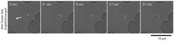
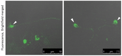
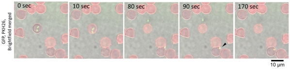
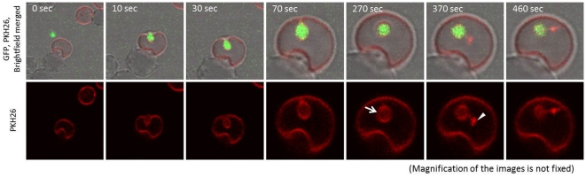
Methodology/principal findings: Gliding motility of B. bovis merozoites was revealed by time-lapse video microscopy. The recorded images revealed that the process included egress of the merozoites from the infected RBC, gliding motility, and subsequent invasion into new RBCs. The gliding motility of B. bovis merozoites was similar to the helical gliding of Toxoplasma tachyzoites. The trails left by the merozoites were detected by indirect immunofluorescence assay using antiserum against B. bovis merozoite surface antigen 1. Inhibition of gliding motility by actin filament polymerization or depolymerization indicated that the gliding motility was driven by actomyosin dependent process. In addition, we revealed the timing of breakdown of the parasitophorous vacuole. Time-lapse image analysis of membrane-stained bovine RBCs showed formation and breakdown of the parasitophorous vacuole within ten minutes of invasion.

Conclusions/significance: This is the first report of the gliding motility of B. bovis. Since merozoites of Plasmodium parasites do not glide on a substrate, the gliding motility of B. bovis merozoites is a notable finding.


Conflict of interest statement

Competing Interests: The authors have declared that no competing interests exist.

Figures

Figure 1
Figure 1. Time-lapse video microscopy of a B. bovis merozoite engaged in gliding.
In vitro cultured IRBCs were placed on a glass slide and their motility documented with confocal laser microscopy. The time elapsed between each frame is indicated in seconds. Gliding of the B. bovis merozoites is characterized as forward movement with the apical end at the front position.
Figure 2
Figure 2. Helical motility of B. bovis merozoites.
In vitro cultured IRBCs were stained with a fluorescent mitochondrial probe (MitoTracker Red) and placed on a glass slide for confocal laser microscopy analysis. The shift in red fluorescence on the merozoites (top to bottom) indicates helical motility of the merozoites. The arrow indicates the direction of merozoite movement.
Figure 3
Figure 3. B. bovis merozoites deposited surface membrane trails during gliding on a solid substrate.
An indirect immunofluorescence antibody assay was performed by using anti-B. bovis MSA-1 mouse antiserum. Arrow heads show the fluorescence from the merozoites.
Figure 4
Figure 4. Time-lapse video microscopy of B. bovis merozoite egress from a RBC, gliding to and invasion of a new RBC.
The arrow head shows merozoite invasion into a new RBC. The red ring-shaped fluorescence around the merozoite indicates the parasitophorous vacuole. The time elapsed between each frame is indicated in seconds.
Figure 5
Figure 5. Time-lapse video microscopy of the formation and breakdown of the parasitophorous vacuole.
To enable monitoring the PV, PKH26 stained fresh RBC was added to the unstained IRBCs. PKH26 fluorescence can be observed around the merozoite just after invasion into the new RBC (arrow). Subsequently, red dot-shaped fluorescence appears (arrow head), and the ring-shaped fluorescence becomes weaker. The time elapsed between each frame is indicated in seconds.

Similar articles

Cited by

References

    1. King CA. Cell motility of sporozoan protozoa. Parasitol Today. 1988;4:315–319. - PubMed
    1. Sibley LD. Intracellular parasite invasion strategies. Science. 2004;304:248–253. - PubMed
    1. Vanderberg JP. Studies on the motility of Plasmodium sporozoites. J Protozool. 1974;21:527–537. - PubMed
    1. Håkansson S, Morisaki H, Heuser J, Sibley LD. Time-lapse video microscopy of gliding motility in Toxoplasma gondii reveals a novel, biphasic mechanism of cell locomotion. Mol Biol Cell. 1999;10:3539–3547. - PMC - PubMed
    1. Wetzel DM, Schmidt J, Kuhlenschmidt MS, Dubey JP, Sibley LD. Gliding motility leads to active cellular invasion by Cryptosporidium parvum sporozoites. Infect Immun. 2005;73:5379–5387. - PMC - PubMed

Publication types