. 2012 Mar;1(1):1-9.
doi: 10.1007/s13669-011-0004-1.

Ovarian Cancer Is an Imported Disease: Fact or Fiction?

Affiliations

Ovarian Cancer Is an Imported Disease: Fact or Fiction?

Elisabetta Kuhn et al. Curr Obstet Gynecol Rep. 2012 Mar.

Abstract

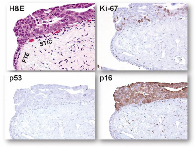
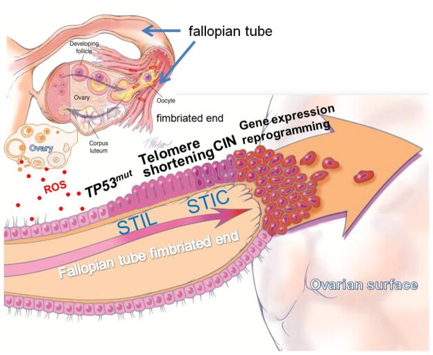
The cell of origin of ovarian cancer has been long debated. The current paradigm is that epithelial ovarian cancer (EOC) arises from the ovarian surface epithelium (OSE). OSE is composed of flat, nondescript cells more closely resembling the mesothelium lining the peritoneal cavity, with which it is continuous, rather than the various histologic types of ovarian carcinoma (serous, endometrioid, and clear cell carcinoma), which have a Müllerian phenotype. Accordingly, it has been argued that the OSE undergoes a process termed "metaplasia" to account for this profound morphologic transformation. Recent molecular and clinicopathologic studies not only have failed to support this hypothesis but also have provided evidence that EOC stems from Müllerian-derived extraovarian cells that involve the ovary secondarily, thereby calling into question the very existence of primary EOC. This new model of ovarian carcinogenesis proposes that fallopian tube epithelium (benign or malignant) implants on the ovary to give rise to both high-grade and low-grade serous carcinomas, and that endometrial tissue implants on the ovary and produces endometriosis, which can undergo malignant transformation into endometrioid and clear cell carcinoma. Thus, ultimately EOC is not ovarian in origin but rather is secondary, and it is logical to conclude that the only true primary ovarian neoplasms are germ cell and gonadal stromal tumors analogous to tumors in the testis. If this new model is confirmed, it has profound implications for the early detection and treatment of "ovarian cancer."


Conflict of interest statement

Disclosure

No potential conflicts of interest relevant to this article were reported.

Figures

Figure 1
Figure 1
Morphologic and molecular genetic features that characterize each major subtype of ovarian carcinoma. The top panels of photomicrographs illustrate their histologic features. The bottom table shows the frequency of sequence mutations of oncogenes and tumor suppressor genes in a heat map. The frequency of mutation is represented by the gradient of colors, from green (low frequency) to red (high frequency). Of note, the case number is too small to be conclusive for PPP2R1A mutations in mucinous tumor. HG, high-grade serous carcinoma; LG, low-grade serous carcinoma; CC, clear cell carcinoma; EM, endometrioid carcinoma; MU, mucinous carcinoma.
Figure 2
Figure 2
The histology and expression of Ki-67, p53, and p16 in a serous tubal intraepithelial carcinoma (STIC). Hematoxylin and eosin (H&E) stained fallopian tube section shows a STIC, composed of enlarged and highly atypical tumor cells, forming a multilayered architecture. No evidence of invasion is noted. In contrast, the adjacent normal fallopian tube epithelium (FTE) contains single-layered, smaller and homogenous epithelial cells. The area was immunostained with antibodies reacting to Ki-67, p53, and p16. The percentage of Ki-67 labeled cells is greater in STIC than in normal fallopian tube epithelium, indicating a higher proliferative activity in STIC. p53 staining is undetectable in STIC because the lesion harbors a truncating mutation in TP53. STIC cells demonstrate diffuse p16 immunoreactivity, suggesting an Rb pathway aberration.
Figure 3
Figure 3
Hypothesis for the tubal origin of ovarian high-grade serous carcinoma (HGSC). We hypothesize that normal fallopian tube epithelium (FTE) is the cell of origin of many “ovarian” HGSCs. Rupture of the dominant follicle at ovulation exposes the underlying ovarian stroma to fimbrial epithelium, which can implant on the ruptured ovarian surface. Inflammation and repair occur in the presence of follicular fluid, which contains a high concentration of reactive oxygen species (ROS, red dots). The increased genotoxic effects may facilitate the selection of TP53 mutations in epithelial cells, which clonally expand. As a result, telomere shortening occurs and enhances underlying chromosomal instability (CIN), creating a repertoire of tumor subclones in serous tubal intraepithelial lesions (STIL), some of which may acquire malignant phenotypes (serous tubal intraepithelial carcinoma, STIC) and exfoliate onto the ovarian and peritoneal surfaces. In summary, FTE may lead to ovarian HGSC by two different mechanisms: (1) normal FTE implants on the ruptured ovarian surface at ovulation, which invaginates to form an inclusion cyst that subsequently undergoes malignant transformation (possibly after a TP53 mutation), or (2) STIC cells implant on the ovary and then form a tumor mass. In both instances, the ovarian HGSC is of tubal origin.

References

    1. Buys SS, Partridge E, Black A, et al. Effect of screening on ovarian cancer mortality: the Prostate, Lung, Colorectal and Ovarian (PLCO) Cancer Screening Randomized Controlled Trial. JAMA. 2011;305:2295–2303. This population-based study analyzed the PLCO cases in an attempt to determine whether CA125 and an imaging study can save the lives of ovarian cancer patients. The study result provides cogent evidence of a lack of effective screening to reduce ovarian cancer deaths. - PubMed
    1. Jemal A, Bray F, Center MM, et al. Global cancer statistics. CA Cancer J Clin. 2011;61:69–90. - PubMed
    1. Fishman DA, Cohen L, Blank SV, et al. The role of ultrasound evaluation in the detection of early-stage epithelial ovarian cancer. Am J Obstet Gynecol. 2005;192:1214–1221. discussion 1221–1212. - PubMed
    1. Kurman RJ, Visvanathan K, Roden R, et al. Early detection and treatment of ovarian cancer: shifting from early stage to minimal volume of disease based on a new model of carcinogenesis. Am J Obstet Gynecol. 2008;198:351–356. - PMC - PubMed
    1. Seidman JD, Cho K, Ronnett BM, et al. Surface epithelial tumors of the ovary. In: Kurman RJ, Ellenson LH, Ronnett BM, editors. Blaustein’s Pathology of the Female Genital Tract. New York: Springer Verlag; 2011. pp. 679–784.