Kambin's Triangle Approach of Lumbar Transforaminal Epidural Injection with Spinal Stenosis
- PMID: 22506212
- PMCID: PMC3309379
- DOI: 10.5535/arm.2011.35.6.833
Kambin's Triangle Approach of Lumbar Transforaminal Epidural Injection with Spinal Stenosis
Abstract
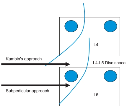
Objective: To compare the short-term effect and advantage of transforaminal epidural steroid injection (TFESI) performed using the Kambin's triangle and subpedicular approaches.
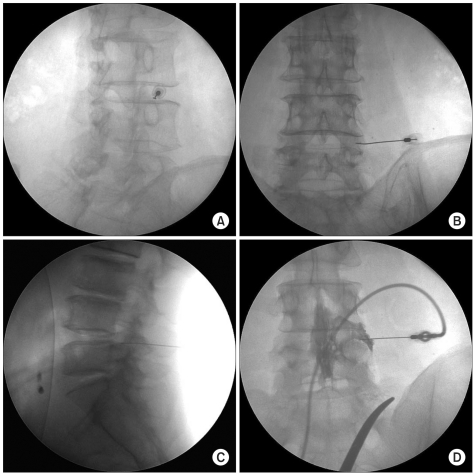
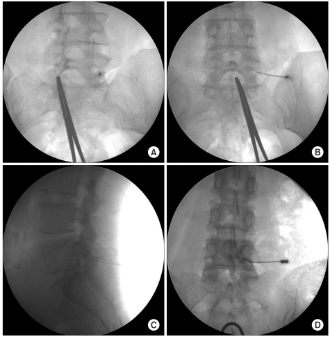
Method: Forty-two patients with radicular pain from lumbar spinal stenosis were enrolled. Subjects were randomly assigned to one of two groups. All procedures were performed using C-arm KMC 950. The frequency of complications during the procedure and the effect of TFESI at 2 and 4 weeks after the procedure between the two groups were compared. Short-term outcomes were measured using a visual numeric scale (VNS) and a five-grade scale. Multiple logistic regression analyses were performed to evaluate the relationship between possible outcome predictors (Kambin's triangle or subpedicular approach, age, duration of symptoms and sex) and the therapeutic effect.
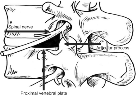
Results: VNS was improved 2 weeks after the injection and continued to improve until 4 weeks in both groups. There were no statistical differences in changes of VNS, effectiveness and contrast spread pattern between these two groups. No correlation was found between the other variables tested and therapeutic effect. Spinal nerve pricking occurred in five cases of the subpedicular and in none of the cases of the Kambin's triangle approach (p<0.05).
Conclusion: The Kambin's triangle approach is as efficacious as the subpedicular approach for short-term effect and offers considerable advantages (i.e., less spinal nerve pricking during procedure). The Kambin's triangle approach maybe an alternative method for transforaminal epidural steroid injection in cases where needle tip positioning in the anterior epidural space is difficult.
Keywords: Kambin's triangle; Lumbar; Stenosis; Transforaminal.
Figures

References
-
- Grubb SA, Lipscomb HJ, Coonrad RW. Degenerative adult onset scoliosis. Spine. 1988;13:241–245. - PubMed
-
- Grubb SA, Lipscomb HJ, Suh PB. Results of surgical treatment of painful adult scoliosis. Spine (Phila Pa 1976) 1994;15:1619–1627. - PubMed
-
- Jackson RP, McManus AC. Radiographic analysis of sagittal plane alignment and balance in standing volunteers and patients with low back pain matched for age,sex, and size. A prospective controlled clinical study. Spine. 1994;19:1611–1161. - PubMed
-
- Rydevik B, Brown MD, Lundborg G. Pathoanatomy and pathophysiology of nerve root compression. Spine. 1984;9:7–15. - PubMed
-
- Olmarker K, Redevik B, Holm S. Edema formation in spinal nerve roots induced by experimental, graded compression. An experimental study on the pig cauda equina with special reference to differences in effects between rapid and slow onset of compression. Spine. 1989;14:569–573. - PubMed
LinkOut - more resources
Full Text Sources
Other Literature Sources