. 2012 Jun 8:4:11.
doi: 10.1186/1758-3284-4-11.

Knockdown of aberrantly expressed nuclear localized decorin attenuates tumour angiogenesis related mediators in oral cancer progression model in vitro

Affiliations

Knockdown of aberrantly expressed nuclear localized decorin attenuates tumour angiogenesis related mediators in oral cancer progression model in vitro

Nyla Dil et al. Head Neck Oncol. .

Expression of concern in

  • Comment: Head and Neck Oncology.
    BioMed Central. BioMed Central. BMC Med. 2014 Feb 5;12:24. doi: 10.1186/1741-7015-12-24. BMC Med. 2014. PMID: 24499430 Free PMC article. Review.

Abstract

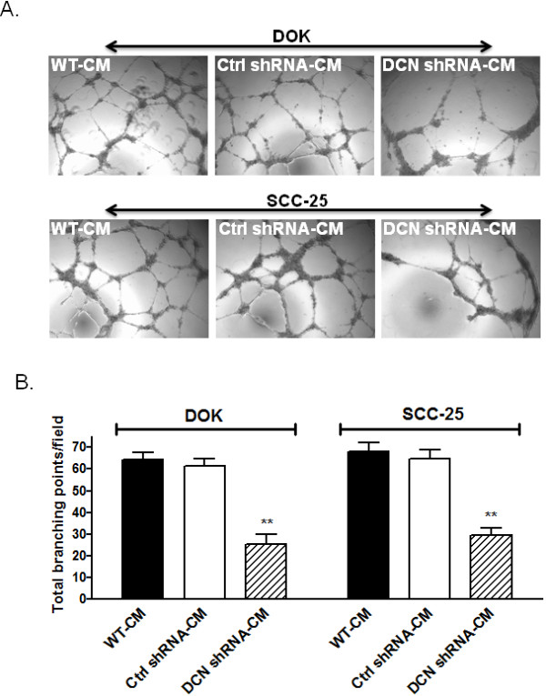
Background: Oral cancer accounts for roughly 3% of cancer cases in the world with about 350,000 newly reported cases annually and a 5-year survival rate of only 50%. Majority of oral cancers are squamous cell carcinomas that originate in the oral mucosal epithelial linings. We have previously shown that in human malignant squamous cells carcinoma (SCC-25) as well as in dysplastic oral keratinocytes (DOK), a small leucine-rich multifunctional proteoglycan decorin is aberrantly expressed and localized in the nucleus where it interacts with nuclear epidermal growth factor receptor (EGFR). Post-transcriptional silencing of nuclear decorin significantly reduced IL-8 and IL8-dependent migration and invasion in these dysplastic and malignant oral epithelia. The objective of this study was to further examine the effects of nuclear decorin silencing on angiogenesis and angiogenesis related mediators in this oral cancer progression cell line model.

Methods: We have used multiplex PCR, western blotting, and in vitro endothelial tube formation assay to study angiogenesis and related pathways in nuclear decorin silenced (stable knockdown) DOK and SCC-25 cells.

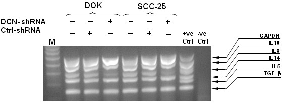
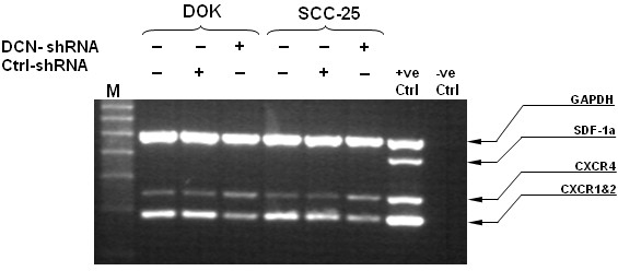
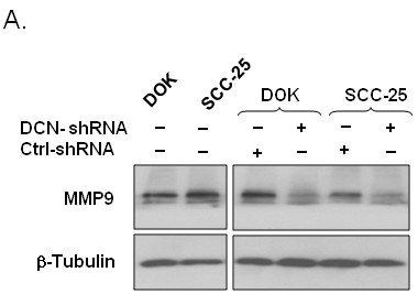
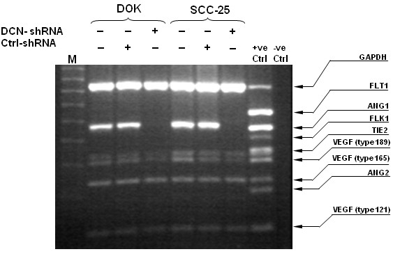
Results: Nuclear decorin knockdown resulted in significant down regulation of IL-8 expression, however IL-10, and TGF-β expression was not affected in either DOK or SCC25 cells as measured by multiplex RT PCR. IL-8 receptor CXCR 1 and 2 expression was slightly lower in nuclear decorin silenced cells indicating a contributing mechanism in previously shown reduced IL-8 mediated migration and invasion phenotype in these cells. IL-8 is known to induce Matrix metalloproteinase 9 (MMP9) which not only plays a role in tumour migration and invasion but also induces angiogenic switch. We found MMP9 to be significantly reduced in nuclear decorin silenced dysplastic and malignant oral epithelia. Other potent angiogenic mediators, VEGF189 and ANG-1 were either significantly reduced or completely abrogated in these cells. Angiogenesis as measured by endothelial tube-like formations of HUVEC cells was reduced by almost 50 percent when HUVECs were incubated in the presence of conditioned medium form nuclear decorin silenced dysplastic and malignant cell lines as compared to respective controls.

Conclusions: Together these results indicate that aberrantly expressed nuclear localized decorin strongly influences angiogenic potential of dysplastic and malignant oral epithelial cells.


Figures

Figure 1
Figure 1
Effect of nuclear decorin silencing on cytokines/chemokines expression in decorin silenced DOK and SCC-25 cells. DOK and SCC-25 cells were stably transfected with decorin-shRNA (DCN-shRNA), or scrambled sequence-shRNA (Ctrl-shRNA) or no transfection control (WT). RNA was extracted and cDNA was subjected to multiplex RT-PCR to detect multiple cytokine transcripts. MPCR products were fractionated electrophoretically on a 2% agarose gel containing 0.5 mg/ml ethidium bromide, visualized under UV light and photographed. The results shown are representative of three independent experiments.
Figure 2
Figure 2
Nuclear decorin silencing affects IL-8 receptor expression in decorin silenced DOK and SCC-25 cells. RNA was extracted from WT, control and decorin silenced DOK and SCC-25 cells and cDNA was subjected to multiplex RT-PCR to detect CXCR1&2 expressions along with other receptors. MPCR products were fractionated electrophoretically on a 2% agarose gel containing 0.5 mg/ml ethidium bromide, visualized under UV light and photographed. The results shown are representative of three independent experiments.
Figure 3
Figure 3
MMP9 expression down regulation in decorin silenced DOK and SCC25 cells. Cell lysates from decorin silenced and control dysplastic and malignant oral epithelia were collected as described in materials and methods and subjected to SDS-PAGE followed by immunoblotting using anti-MMP9 and anti-β-tubulin antibodies. Data shown from one representative experiment of three.
Figure 4
Figure 4
VEGF and ANG-1 expression modulation in decorin silenced DOK and SCC25 cells. RNA was extracted from WT, control and decorin silenced DOK and SCC-25 cells and cDNA was subjected to multiplex RT-PCR to detect the expression of angiogenesis related mediators. MPCR products were fractionated electrophoretically on a 2% agarose gel containing 0.5 mg/ml ethidium bromide, visualized under UV light and photographed. The results shown are representative of three independent experiments.
Figure 5
Figure 5
Overall influence of nuclear decorin silencing on angiogenic capacity of DOK and SCC-25 cell derived factors; as assessed on human endothelial cellsin vitro.A, HUVECs (3X10⁴) were plated on ECM coated wells along with 150 ul of either WT, control or decorin silenced conditioned medium (CM). Endothelial tube formation was assessed after incubating at 37°C for 18 h, using a light microscope. Three microscopic fields were selected at random and photographed at 200x magnification. B, Tube formation ability was quantified by counting the total number of cell clusters and branching under three fields per well. The results are expressed as mean of branching compared with the control groups. Each experiment was performed at least twice. Numbers represent mean ± SD of three individual experiments. ** p <;0.01, *** p <;0.001 compared to respective controls.

References

    1. Cooper JS, Porter K, Mallin K, Hoffman HT, Weber RS, Ang KK, Gay EG, Langer CJ. National Cancer Database report on cancer of the head and neck: 10-year update. Head Neck. 2009;31:748–758. doi: 10.1002/hed.21022. - DOI - PubMed
    1. Jemal A, Siegel R, Ward E, Hao Y, Xu J, Murray T, Thun MJ. Cancer statistics. CA Cancer J Clin. 2008;58:71–96. doi: 10.3322/CA.2007.0010. - DOI - PubMed
    1. Parkin DM, Bray F, Ferlay J, Pisani P. Global cancer statistics, 2002. CA Cancer J Clin. 2005;55:74–108. doi: 10.3322/canjclin.55.2.74. - DOI - PubMed
    1. de Camargo Cancela M, Voti L, Guerra-Yi M, Chapuis F, Mazuir M, Curado MP. Oral cavity cancer in developed and in developing countries: population-based incidence. Head Neck. 2010;32:357–367. - PubMed
    1. Hanahan D, Folkman J. Patterns and emerging mechanisms of the angiogenic switch during tumorigenesis. Cell. 1996;86:353–364. doi: 10.1016/S0092-8674(00)80108-7. - DOI - PubMed

Publication types

MeSH terms