

Case Reports
doi: 10.1186/1751-0147-54-24.
Magnetic resonance and computed tomography imaging of a carotid body tumor in a dog
Affiliations
- PMID: 22507757
- PMCID: PMC3403989
- DOI: 10.1186/1751-0147-54-24
Item in Clipboard
Case Reports
Magnetic resonance and computed tomography imaging of a carotid body tumor in a dog
Acta Vet Scand.
.
Abstract
A 5-year-old castrated male Labrador Retriever was presented to a referring veterinarian for a swelling in the neck region. Based on the results of histopathology, a carotid body tumor, was diagnosed. The dog was referred to a medical imaging unit for further staging and follow up. This report describes the magnetic resonance (MR) and computed tomographic (CT) appearance of a carotid body tumor.
Figures

Lymph node metastasis of the carotid body tumor in a lymph node (c), composed of small nests of large pleiomorphic polygonal or oval cells (arrow) with a large round nucleus with coarse chromatin and inconspicuous nucleoli, and a variable amount of granular, slightly basophilic cytoplasm (H&H stain; bar=80m). Also notice the mild cytoplasmic staining with anti-chromogranin A antibodies (inset right, b, immunohistochemical stain with anti-chromogranine A, bar=50m) and strong cytoplasmic staining with anti-vimentin antibodies (inset left, a, immunohistochemical stain; anti-vimentin, bar=50m)

Sagittal T2W SE image of the left cervical region. A heterogenous, hyperintense mass (carotid body tumor) (arrow) with multiple signal voids is visible caudal from the bulla (b) to the caudal part of the atlas (C1). A lobulated, elongated more homogeneous mass (medial retropharyngeal lymph node) (asterisk) is visible streching out to the caudal part of C4. The CCA (cca) is visible laterally from this mass.

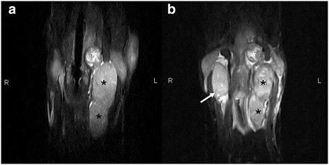
Transverse T2W SE images at the level of the caudal part of the bulla (a), cranial part of the atlas (b) and the mid part of the axis (c). A heterogenous, hyperintense mass (carotid body tumor) (black asterisk) is visible on the left side, with multiple signal voids of vessels. The mass splays the ICA more medially (large arrow) and the ECA more laterally (small arrow). Laterally a homogeneous, hyperintense encapsulated mass (medial retropharyngeal lymph node) (white asterisk) is visible. The mass is located between the CCA (arrowhead) and the mandibular gland (m) (panel b) and more caudally between the CCA and the m. sternocephalicus (s).

Dorsal reformatted CT images of the cervical region. Images are displayed in a soft tissue window. The pre-contrast image (a) shows a well defined soft-tissue mass (carotid body tumor) (white asterisk) splaying the ICA medially (large arrow) and the ECA laterally (small arrow). Also a more lobulated, elongated, encapsulated soft- tissue mass (medial retropharyngeal lymph node) (black asterisk) with areas of calcification is visible on the left side, laterally from the CCA (arrowhead). On the right side a singular soft-tissue mass (medial retropharyngeal lymph node) (red asterisk) is visible. On the post-contrast image (b) the mass shows a heterogenous intense contrast enhancement with areas of low density (necrosis). Both the elongated mass on the left side and the singular mass on the right side have a contrast uptaking capsula. The splaying of the ICA and ECA by the mass is clearly visible.
Dorsal STIR images of the cervical region during the first examination (a) and the follow up examination (b). The primary heterogenous mass (carotid body tumor) (white asterisk) and the secondary lobulated, homogeneous mass (medial retropharyngeal lymph node) (black asterisk) appear to be decreased in size (panel a compared to panel b). A secondary, homogeneous, encapsulated medial retropharyngeal lymph node (arrow) is visible on the right side (panel b).

Postmortem image of the left cranial cervical region. The carotid body tumor (asterisk) is localized just craniomedial of the ECA (arrow). CCA is visible (two arrows).
Similar articles
-
CT AND MRI FEATURES OF CAROTID BODY PARAGANGLIOMAS IN 16 DOGS.Vet Radiol Ultrasound. 2015 Jul-Aug;56(4):374-83. doi: 10.1111/vru.12254. Epub 2015 Apr 5. Vet Radiol Ultrasound. 2015. PMID: 25846946
-
Imaging diagnosis--computed tomographic, surgical, and histopathologic characteristics of an infiltrative angiolipoma in a dog.Vet Radiol Ultrasound. 2015 May-Jun;56(3):E31-5. doi: 10.1111/vru.12178. Epub 2014 May 22. Vet Radiol Ultrasound. 2015. PMID: 24852319
-
Canine malignant carotid body tumor.J Am Vet Med Assoc. 1970 Mar 1;156(5):606-10. J Am Vet Med Assoc. 1970. PMID: 5461696 No abstract available.
-
Carotid body tumor: a case report and literature review.J Radiol Case Rep. 2019 Aug 31;13(8):19-30. doi: 10.3941/jrcr.v13i8.3681. eCollection 2019 Aug. J Radiol Case Rep. 2019. PMID: 31558967 Free PMC article. Review.
-
Intracranial neoplasia.Clin Tech Small Anim Pract. 1999 May;14(2):112-23. doi: 10.1016/S1096-2867(99)80009-7. Clin Tech Small Anim Pract. 1999. PMID: 10361361 Review.
Cited by
-
Intra-arterial versus intra venous contrast-enhanced computed tomography of the equine head.BMC Vet Res. 2016 Jan 7;12:6. doi: 10.1186/s12917-016-0632-9. BMC Vet Res. 2016. PMID: 26739315 Free PMC article.
-
An atypical case of recurrent carotid body carcinoma in a young adult dog: Histopathological, immunohistochemical and electron microscopic study.J Vet Med Sci. 2017 Apr 5;79(4):714-718. doi: 10.1292/jvms.16-0501. Epub 2017 Feb 27. J Vet Med Sci. 2017. PMID: 28239052 Free PMC article.
References
-
- Hayes HM, Sass B. Chemoreceptor neoplasia: a study of the epidemiological features of 357 canine cases. Zentralbl Veterinarmed (A) 1988;35(6):401–408. - PubMed
-
- Noszczyk-Nowak A, Nowak M, Paslawska U, Atamaniuk W, Nicpon J. Cases with manifestation of chemodectoma diagnosed in dogs in Department of Internal Diseases with Horses, Dogs and Cats Clinic, Veterinary Medicine Faculty, University of Environmental and Life Sciences, Wroclaw. Poland. Acta Vet Scan. 2010;52:35. - PMC - PubMed
-
- Dean MJ, Straffuss AC. Carotid body tumors in the dog: a review and report of four cases. J Am Vet Med Assoc. 1975;166(10):1003–1006. - PubMed
Publication types
MeSH terms
LinkOut - more resources
Full Text Sources
Medical
