. 2012 Sep;22(9):1390-401.
doi: 10.1038/cr.2012.61. Epub 2012 Apr 17.

Cep57, a NEDD1-binding pericentriolar material component, is essential for spindle pole integrity

Affiliations

Cep57, a NEDD1-binding pericentriolar material component, is essential for spindle pole integrity

Qixi Wu et al. Cell Res. 2012 Sep.

Abstract

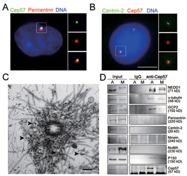
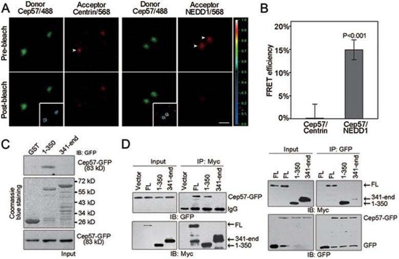
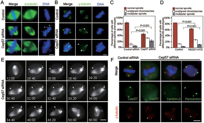
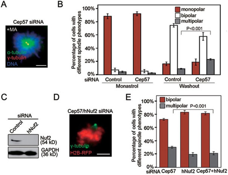
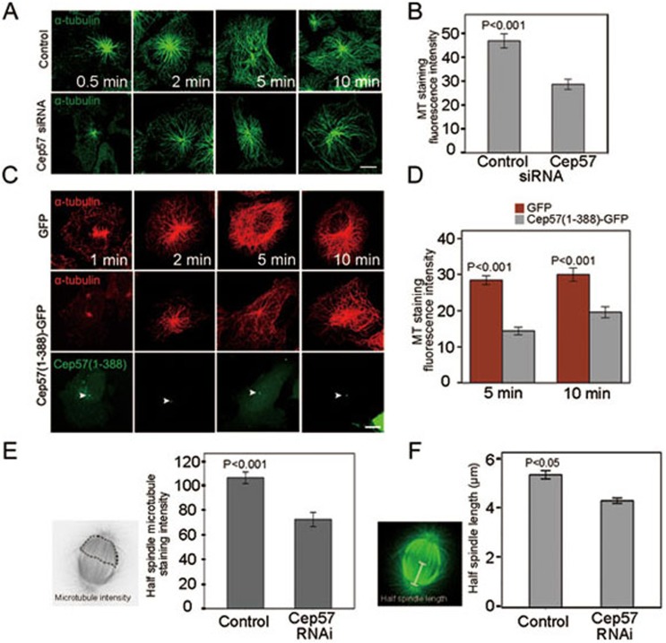
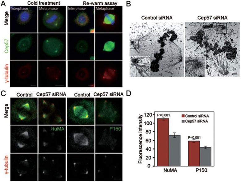
Formation of a bipolar spindle is indispensable for faithful chromosome segregation and cell division. Spindle integrity is largely dependent on the centrosome and the microtubule network. Centrosome protein Cep57 can bundle microtubules in mammalian cells. Its related protein (Cep57R) in Xenopus was characterized as a stabilization factor for microtubule-kinetochore attachment. Here we show that Cep57 is a pericentriolar material (PCM) component. Its interaction with NEDD1 is necessary for the centrosome localization of Cep57. Depletion of Cep57 leads to unaligned chromosomes and a multipolar spindle, which is induced by PCM fragmentation. In the absence of Cep57, centrosome microtubule array assembly activity is weakened, and the spindle length and microtubule density decrease. As a spindle microtubule-binding protein, Cep57 is also responsible for the proper organization of the spindle microtubule and localization of spindle pole focusing proteins. Collectively, these results suggest that Cep57, as a NEDD1-binding centrosome component, could function as a spindle pole- and microtubule-stabilizing factor for establishing robust spindle architecture.


Figures

Figure 1
Figure 1
Cep57 is a PCM component. (A, B) Immunostaining of Cep57 (green, A; red, B), pericentrin (red, A) or centrin-2 (green, B) in HeLa cells. Nuclear DNA was stained with DAPI (blue) in all figures of this paper. Boxed regions, close-up of the centrosome. Bar, 5 μm. (C) Immunoelectron microscopy images of HeLa cell centrosome. Arrowheads indicate 5 nm gold particles. Bar, 100 nm. (D) Cep57 is associated with γ-tubulin ring complex components. Asynchronized (A) or metaphase-arrested (M) HeLa cell lysates were immunoprecipitated with anti-Cep57 antibody or non-specific rabbit IgG. The inputs (left) and immunoprecipitants (right) were immunoblotted with the indicated antibodies.
Figure 2
Figure 2
Cep57 binds to the N-terminus of NEDD1. (A, B) Immunostaining of Cep57 (Alexa Fluro 488) and centrin-2 or NEDD1 (Alexa Fluro 568) in HeLa cells, which were subjected to acceptor photobleaching. Representative images show the pre- and post-photobleaching states of the centrosome. Insets show the FRET intensities encoded by using the scale bar on the right. Bar, 1 μm. The graph shows the FRET efficiencies. n = 10. Error bars represent mean ± SEM. (C) Lysates of 293T cells expressing Cep57-GFP were incubated with glutathione-agarose beads coated with GST or GST-NEDD1 fusion protein. The inputs and the proteins bound to the beads were immunoblotted with anti-GFP antibody. (D) Co-IP with anti-Myc and anti-GFP antibodies were performed in lysates of 293T cells coexpressing Cep57-GFP and Myc vector or Myc-NEDD1 constructs, followed by immunoblotting with the indicated antibodies. FL, Myc-NEDD1 full-length. 1-350, Myc-NEDD1 (1-350). 341-end, Myc-NEDD1 (341-end).
Figure 3
Figure 3
The centrosome localization of Cep57 requires its interaction with NEDD1. (A) Representative phenotypes of Cep57-GFP-transfected cells. Bar, 10 μm. (B) The graph shows the proportion of cells with indicated phenotypes (n = 3; > 80 cells per experiment). Error bars represent mean ± SEM. (C) At 72 h after Cep57 siRNA transfection, the protein levels of Cep57 and NEDD1 in HeLa cells were examined by immunoblotting with the indicated antibodies. (D) At 65 h after NEDD1 siRNA transfection, HeLa cells were subjected to DMSO or MG132 treatment. Protein expression of Cep57 and NEDD1 was examined by immunoblotting with the indicated antibodies. (E-I) NEDD1 RNAi results in the loss of Cep57 from the centrosome. (E, F, H) Immunofluorescence images of HeLa cells treated with the indicated siRNAs. NEDD1 (green), Cep57 (red). (G, I) Quantification of the fluorescence intensities within the centrosome (n = 3; > 60 cells per experiment). Error bars represent mean ± SEM. (J, K) Immunostaining of Cep57 (green) in HeLa cells transfected with NEDD1 truncates (red). Arrows and arrowheads indicate the centrosomes in untransfected and transfected cells, respectively. Bars, 5 μm.
Figure 4
Figure 4
Depletion of Cep57 causes spindle assembly defects and PCM fragmentation. (A, B) Immunostaining of α-tubulin (green, A) or γ-tubulin (green, B) in HeLa cells. Arrowheads indicate unaligned chromosomes (A) or extra spindle poles (B). (C, D) Quantification of different spindle phenotypes in synchronized metaphase HeLa cells after siRNA treatment (C) or NEDD1-NTD transfection (D) (n = 3; > 100 cells per experiment). Error bars represent mean ± SEM. (E) Representative time-lapse images of the mitotic progression of HeLa cells cotransfected with Cep57 siRNAs and α-tubulin-pCAsalGFP of Supplementary information, Movie S2. Arrowheads indicate a budded pole. Arrows indicate an unfocused spindle pole. (F) Immunostaining of metaphase siRNA-transfected HeLa cells for Cep57, centrin-2 or pericentrin (green) and γ-tubulin (red). Arrowheads indicate extra spindle poles.
Figure 5
Figure 5
Spindle forces are responsible for the formation of multipolar spindles induced by Cep57 depletion. (A, B) Immunofluorescence images of MA-treated Cep57-depleted cells with α-tubulin (green) and γ-tubulin (red) staining (A). The proportion of mitotic cells with different phenotypes was quantified after MA treatment or MA washout (n = 3; > 200 cells per experiment). Error bars represent mean ± SEM. (C) At 72 h after siRNA transfection, levels of hNuf2 in HeLa cells were examined by immunoblotting. (D, E) Representative images of Cep57 and hNuf2 siRNA-transfected cells with γ-tubulin (green) staining (D). H2B-RFP was used as a transfection marker. Bars, 5 μm. The proportion of mitotic cells with different phenotypes was quantified (n = 3; > 100 cells per experiment). Error bars represent mean ± SEM.
Figure 6
Figure 6
Cep57 functions in spindle microtubule assembly. (A, B) CHO cells were transfected with pSUPER-RFP or pSUPER-RFP-Cep57 and treated with nocodazole. After nocodazole washout, the cells were immunostained for α-tubulin (green; A). The graph shows the results of the microtubule regrowth assay at 5 min. Quantification of tubulin immunofluorescence intensities in cells (n = 3; >100 cells per experiment). Error bars represent mean ± SEM. (C, D) GFP (upper panels) and Cep57 (1-388)-GFP (middle and lower panels) expressing cells were subjected to a microtubule recovery assay. Arrowheads indicate the centrosome localization of Cep57 (1-388)-GFP. Bar, 10 μm. The graph shows the results of the microtubule regrowth assay at 5 and 10 min (n = 3; > 100 cells per experiment). Error bars represent mean ± SEM. (E) Quantification of spindle microtubule intensity indicated by α-tubulin staining in siRNA-treated cells (n = 3; > 75 cells per experiment). Error bars represent mean ± SEM. (F) Half spindle lengths indicated by α-tubulin staining were measured in control and Cep57-depleted cells (n = 3; > 60 cells per experiment). Error bars represent mean ± SEM.
Figure 7
Figure 7
Cep57 binds spindle microtubules and contributes to spindle microtubule organization. (A) HeLa cells in interphase and mitosis were incubated on ice for 1 h and then warmed to 37 °C for 10 min. The recovery state was studied by immunofluorescence using anti-Cep57 (green) and γ-tubulin (red) antibodies. (B) Electron microscopy images of the representative control and Cep57 RNAi spindle. Insets show high magnification images of spindle poles. Bars, 1 μm. (C, D) HeLa cells treated with siRNAs were stained for NuMA (green), P150 (green) and γ-tubulin (red). Bars, 5 μm. Fluorescence intensities in bipolar spindles were measured within a half spindle (D, n = 3; > 100 cells per experiment). Error bars represent mean ± SEM.
Figure 8
Figure 8
Schematic model highlighting the role of Cep57 in robust spindle architecture establishment. Cep57 acts as a microtubule and PCM stabilization factor at the centrosome and along the spindle microtubules. The loss of Cep57 disrupts the balance of forces within the spindle poles and spindle microtubule organization, which in turn results in disorganized spindle morphology, including multipolar spindles and unaligned chromosomes.

Similar articles

Cited by

References

    1. Blagden SP, Glover DM. Polar expeditions – provisioning the centrosome for mitosis. Nat Cell Biol. 2003;5:505–511. - PubMed
    1. Zheng Y, Wong ML, Alberts B, Mitchison T. Nucleation of microtubule assembly by a gamma-tubulin-containing ring complex. Nature. 1995;378:578–583. - PubMed
    1. Palazzo RE, Vogel JM, Schnackenberg BJ, Hull DR, Wu X. Centrosome maturation. Curr Top Dev Biol. 2000;49:449–470. - PubMed
    1. Thein KH, Kleylein-Sohn J, Nigg EA, Gruneberg U. Astrin is required for the maintenance of sister chromatid cohesion and centrosome integrity. J Cell Biol. 2007;178:345–354. - PMC - PubMed
    1. Oshimori N, Ohsugi M, Yamamoto T. The Plk1 target Kizuna stabilizes mitotic centrosomes to ensure spindle bipolarity. Nat Cell Biol. 2006;8:1095–1101. - PubMed

Publication types