. 2012 Jun 7;4(11):3567-76.
doi: 10.1039/c2nr30366c. Epub 2012 Apr 16.

Development of viral nanoparticles for efficient intracellular delivery

Affiliations

Development of viral nanoparticles for efficient intracellular delivery

Zhuojun Wu et al. Nanoscale. .

Abstract

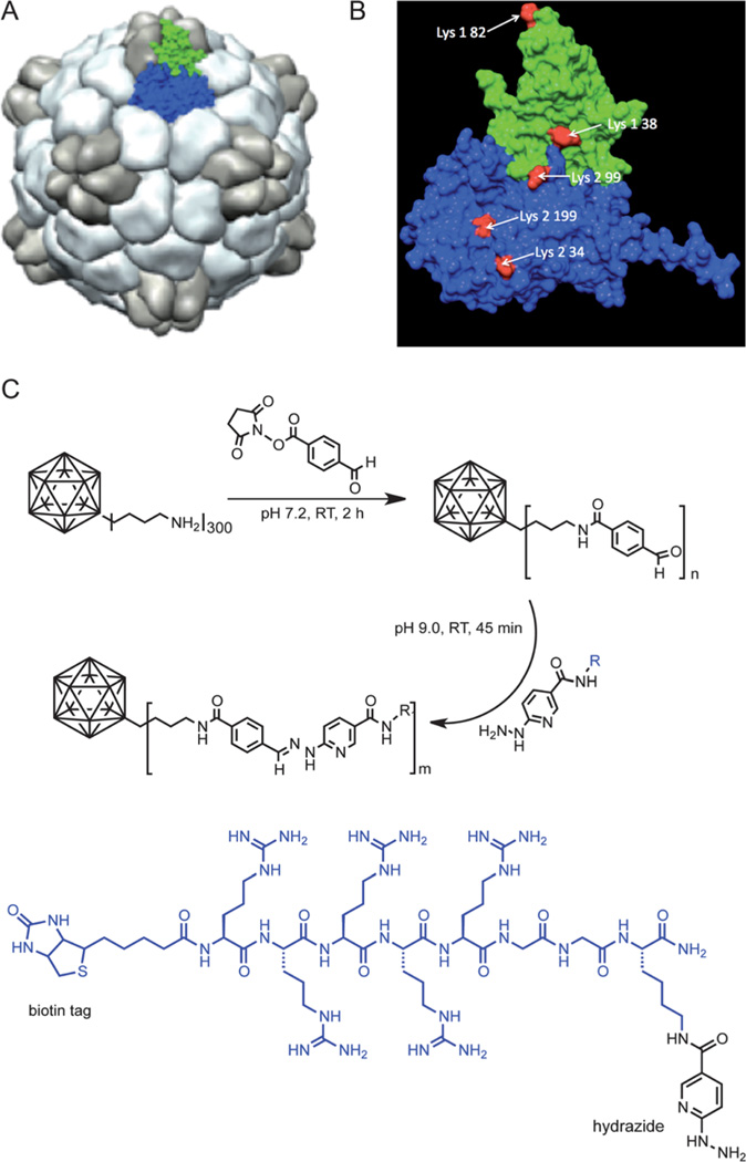
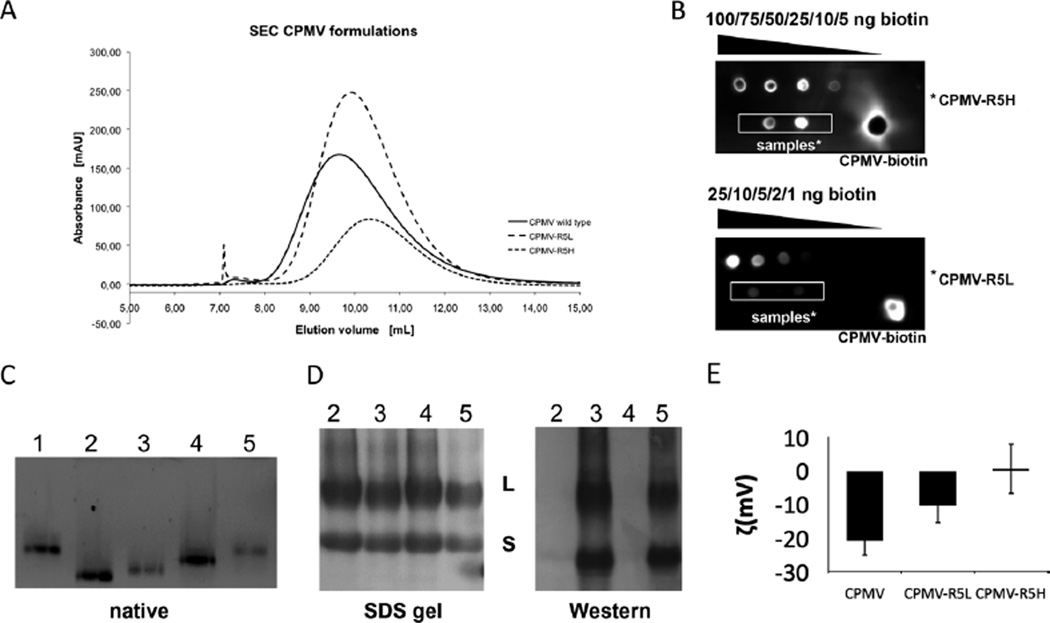
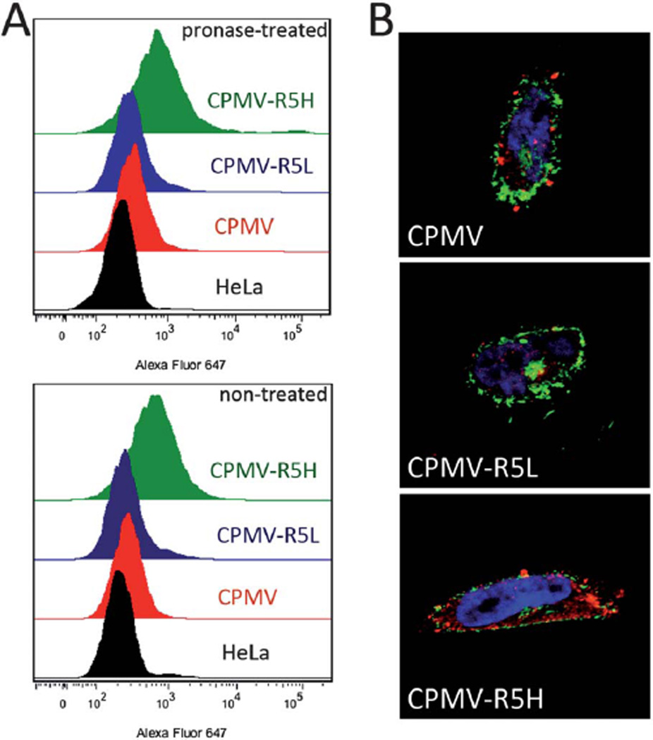
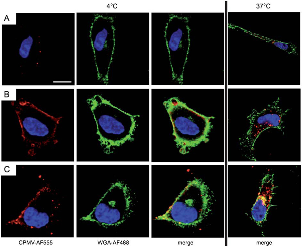
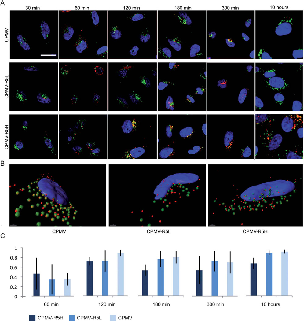
Viral nanoparticles (VNPs) based on plant viruses such as Cowpea mosaic virus (CPMV) can be used for a broad range of biomedical applications because they present a robust scaffold that allows functionalization by chemical conjugation and genetic modification, thereby offering an efficient drug delivery platform that can target specific cells and tissues. VNPs such as CPMV show natural affinity to cells; however, cellular uptake is inefficient. Here we show that chemical modification of the CPMV surface with a highly reactive, specific and UV-traceable hydrazone linker allows bioconjugation of polyarginine (R5) cell penetrating peptides (CPPs), which can overcome these limitations. The resulting CPMV-R5 particles were taken up into a human cervical cancer cell line (HeLa) more efficiently than native particles. Uptake efficiency was dependent on the density of R5 peptides on the surface of the VNP; particles displaying 40 R5 peptides per CPMV (denoted as CPMV-R5H) interact strongly with the plasma membrane and are taken up into the cells via an energy-dependent mechanism whereas particles displaying 10 R5 peptides per CPMV (CPMV-R5L) are only slowly taken up. The fate of CPMV-R5 versus native CPMV particles within cells was evaluated in a co-localization time course study. It was indicated that the intracellular localization of CPMV-R5 and CPMV differs; CPMV remains trapped in Lamp-1 positive endolysosomes over long time frames; in contrast, 30-50% of the CPMV-R5 particles transitioned from the endosome into other cellular vesicles or compartments. Our data provide the groundwork for the development of efficient drug delivery formulations based on CPMV-R5.


Figures

Fig. 1
Fig. 1
(A) Structure of CPMV; surface rendered model highlighting the asymmetric unit consisting of the S protein (green) and L protein (blue). (B) CPMV asymmetric unit; reactive Lys side chains are highlighted in red. (C) Bioconjugation of CPMV with 4FB followed by reaction with R5 peptide.
Fig. 2
Fig. 2
Characterization of CPMV labeling with the biotinylated R5 peptide. (A) Size exclusion chromatography of wild-type CPMV, CPMV–R5L and CPMV–R5Hat 280 nm. (B) ECL dot blot of purified CPMV particles. The number of biotin labels per particle was determined using standardized biotin concentrations and Chemidoc XRS software. (C) Native gel electrophoresis of intact CPMV particles (10 µg) using a 0.8% (w/v) agarose gel. Particles were visualized under UV light. Lane 1 = CPMV, 2 = CPMV–4FB, 3 = CPMV–R5H, 4 = CPMV–PFB, 5 = CPMV–R5L. (D) SDS–PAGE of CPMV particles (10 µg) using a 4–12% Bis-Tris gel and western blotting using streptavidin–alkaline phosphatase to detect the N-terminal biotin tag of the R5 peptide. (E) Zeta potential of CPMV wild type, CPMV–R5L and CPMV–R5H formulations.
Fig. 3
Fig. 3
Uptake of VNPs into human cervical cancer cells (HeLa). (A) Evaluation of particle uptake by flow cytometry. Cells were either untreated or treated with 0.1% (w/v) pronase for 3 h. Experiments were conducted using 105 CPMV particles per cell and repeated at least twice; triplicate samples were analyzed, such that 10 000 events were recorded. (B) Fluorescence confocal microscopy of HeLa cells and CPMV formulations (red). Cell membrane is labeled with wheat germ agglutinin (green), and nucleus is labeled with DAPI (blue). Single plane images were analyzed using ImageJ.
Fig. 4
Fig. 4
Temperature/energy-dependent uptake of CPMV formulations. Confocal microscopy of HeLa cells and CPMV formulations (red). The cell membrane is labeled with wheat germ agglutinin (green), and the nucleus is labeled with DAPI (blue). Scale bar = 10 µm. Cells were incubated for 3 h with 106 VNPs per cell at 4 °C (left) or 37 °C (right). (A) CPMV, (B) CPMV–R5L, (C) CPMV–R5H. Imaging was performed using a Biorad 2100 confocal microscope with a 60× oil objective. Images were analyzed using ImageJ.
Fig. 5
Fig. 5
Subcellular fate of CPMV formulations in HeLa cells studied by confocal microscopy. CPMV formulations (red), endolysosomes are labeled with Lamp-1 (green), and nuclei are labeled with DAPI (blue). The overlay of endolysosomes and VNP signals is shown in yellow. (A) Time course and translocation of CPMV formulation over 10 hours. Scale bar = 10 µm. Images were analyzed using ImageJ. (B) Three-dimensional reconstruction of z-sectional data at time point 120 min. Images were recorded at a step size of 0.3 µm. Data were reconstructed using Imaris software. Scale bar = 1 µm. (C) Colocalization analysis of eight representative cells for each CPMV formulation showing the percentage of total internalized CPMV particles colocalizing with the endolyosomes, error bars represent the averages values of standard deviations of all analyzed cells. Co-localization analysis was performed using LSM Examiner software.

Similar articles

Cited by

References

    1. Steinmetz NF. Nanomedicine. 2010;349 - PMC - PubMed
    2. Huo Q, Liu J, Wang LQ, Jiang Y, Lambert TN, Fang E. J. Am. Chem. Soc. 2006;128:6447. - PubMed
    3. Alexiou C, Schmid RJ, Jurgons R, Kremer M, Wanner G, Bergemann C, Huenges E, Nawroth T, Arnold W, Parak FG. Eur. Biophys. J. 2006;35:446. - PubMed
    4. Dutta T, Agashe HB, Garg M, Balakrishnan P, Kabra M, Jain NK. J. Drug Targeting. 2007;15:89. - PubMed
    5. Zhang L, Gu FX, Chan JM, Wang AZ, Langer RS, Farokhzad OC. Clin. Pharmacol. Ther. 2008;83:761. - PubMed
    1. Jain KK. Med. Princ. Pract. 2008;17:89. - PubMed
    1. Mohanraj V, Chen Y. J. Pharm. Res. 2006;5:561.
    1. Sanvicens N, Marco MP. Trends Biotechnol. 2008;26:425. - PubMed
    2. Torchilin VP. Adv. Drug Delivery Rev. 2006;58:1532. - PubMed
    1. Razis ED, Fountzilas G. Ann. Oncol. 2001;12:593. - PubMed

Publication types