. 2012 Apr 16;197(2):209-17.
doi: 10.1083/jcb.201111115.

The dynamin-like GTPase Sey1p mediates homotypic ER fusion in S. cerevisiae

Affiliations

The dynamin-like GTPase Sey1p mediates homotypic ER fusion in S. cerevisiae

Kamran Anwar et al. J Cell Biol. .

Abstract

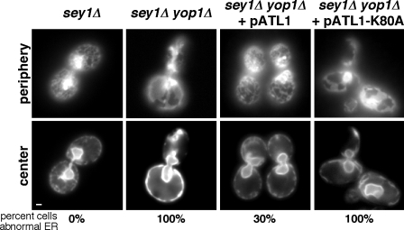
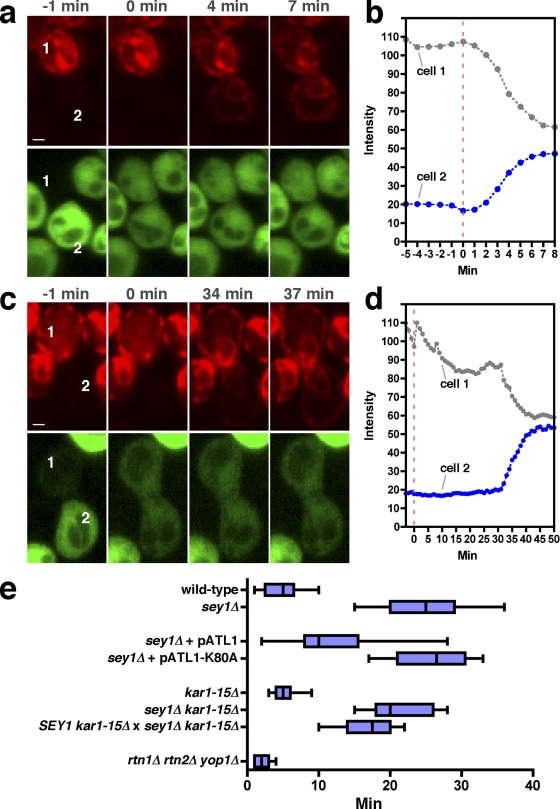
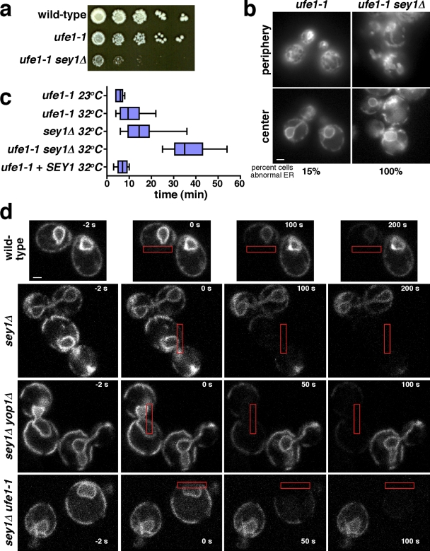
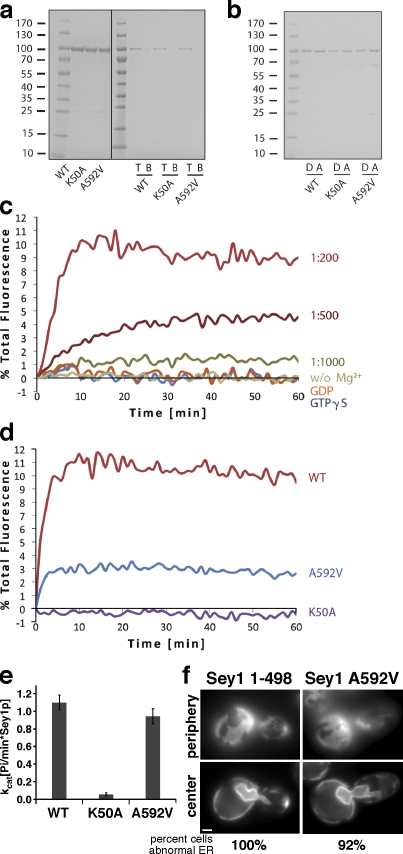
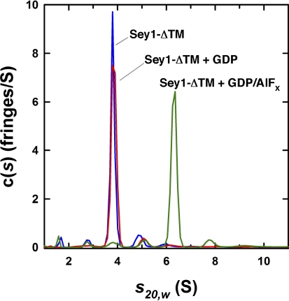
The endoplasmic reticulum (ER) forms a network of tubules and sheets that requires homotypic membrane fusion to be maintained. In metazoans, this process is mediated by dynamin-like guanosine triphosphatases (GTPases) called atlastins (ATLs), which are also required to maintain ER morphology. Previous work suggested that the dynamin-like GTPase Sey1p was needed to maintain ER morphology in Saccharomyces cerevisiae. In this paper, we demonstrate that Sey1p, like ATLs, mediates homotypic ER fusion. The absence of Sey1p resulted in the ER undergoing delayed fusion in vivo and proteoliposomes containing purified Sey1p fused in a GTP-dependent manner in vitro. Sey1p could be partially replaced by ATL1 in vivo. Like ATL1, Sey1p underwent GTP-dependent dimerization. We found that the residual ER-ER fusion that occurred in cells lacking Sey1p required the ER SNARE Ufe1p. Collectively, our results show that Sey1p and its homologues function analogously to ATLs in mediating ER fusion. They also indicate that S. cerevisiae has an alternative fusion mechanism that requires ER SNAREs.


Figures

Figure 1.
Figure 1.
ATL1 can functionally replace Sey1p in yeast. Cells with the indicated genotypes expressing Sec63-GFP were visualized live, focusing on either the center plane or the periphery of the cells. Some cells also contained a plasmid encoding ATL1 or ATL1-K80A, a mutant defective in GTP-binding. Bar, 1 µm.
Figure 2.
Figure 2.
ER–ER fusion is delayed in cells missing Sey1p. (a) Wild-type cells of opposite mating types expressing either ss-RFP-HDEL or cytosolic GFP were mixed, placed on an agarose pad, and imaged at 1-min intervals. Selected images from the time-lapse video are shown. Time 0 is the first image taken after cell fusion, as indicated by GFP in both cells. The two cells of the analyzed mating pair are labeled 1 and 2. Bar, 1 µm. (b) Quantification of the RFP signal of cells 1 and 2 in a. (c) As in a, except that sey1Δ cells were used. (d) Quantification of the RFP signal of cells 1 and 2 in c. (e) A box and whiskers plot of the amount of time between cell fusion and ER fusion during mating. Top and bottom ends of the boxes represent the 75th and 25th percentiles, and whiskers indicate the maximum and minimum values. The median is depicted with a solid line (n = 15–25 from at least three independent experiments). Cells were mated at 23°C. Unless indicated, strains with identical genotypes were mated to each other.
Figure 3.
Figure 3.
Sey1p-independent ER–ER fusion requires the ER SNARE Ufe1p. (a) Serial 10-fold dilutions of cells with the indicated genotypes were spotted onto YPD plates and incubated at 23°C for 5 d. (b) Cells with the indicated genotypes expressing Sec63-GFP were grown at 23°C and visualized live, focusing on either the center plane or the periphery of the cells. (c) A box and whiskers plot of the amount of time between cell fusion and ER fusion when cells with identical genotypes were mated with each other. Top and bottom ends of the boxes represent the 75th and 25th percentiles, and whiskers indicate the maximum and minimum values. The median is depicted with a solid line (n = 12–16 from at least three independent experiments). Cells were grown at 23°C and either mated at this temperature or shifted to 32°C immediately before mating. Where indicated (+ SEY1), the strains contained a plasmid that overexpressed Sey1p. (d) Strains expressing Sec63-GFP were visualized live in growth medium, and images were taken at 2-s intervals. Beginning at time 0, the area in the red rectangle was bleached every 2 s. Selected images from the time-lapse video are shown. All strains except sey1Δ ufe1-1 were grown at 30°C. The sey1Δ ufe1-1 cells were grown at 23°C and then shifted to 37°C for 30 min. Bars, 1 µm.
Figure 4.
Figure 4.
Sey1p-mediated membrane fusion in vitro. (a) Wild-type (WT) Sey1p or the indicated mutants were purified (left blot shows a Coomassie-stained SDS gel) and reconstituted into proteoliposomes. Flotation in a sucrose gradient (right) shows efficient reconstitution of the proteins (T, top fraction; B, bottom fraction). The black line indicates that intervening lanes have been spliced out. (b) Donor (D) and acceptor (A) proteoliposomes containing WT Sey1p or the indicated mutants were analyzed by SDS-PAGE and Coomassie staining. (c) WT Sey1p was reconstituted at the indicated protein to lipid ratios into proteoliposomes containing fluorescent lipids at quenching concentrations or unlabeled lipids (donor and acceptor vesicles, respectively). Donor and acceptor proteoliposomes were mixed at a ratio of 1:3 (600 µM of total lipid) and incubated for 10 min at 37°C before the addition of 1 mM GTP. The increase in fluorescence caused by lipid mixing was followed at 1-min intervals in a fluorescence plate reader. Control reactions were performed in the absence of Mg2+ or in the presence of GDP or GTPγS instead of GTP. (d) Fusion with WT Sey1p was compared with that of the indicated Sey1p mutants. Mutant Sey1K50A has a mutation in the phosphate-binding loop (P-loop) of the active site, and mutant A592V is analogous to one in a plant homologue that causes ER morphology defects. (e) GTPase activity of WT and the indicated Sey1p mutants. Error bars show means ± SD. (f) A sey1Δ yop1Δ strain expressing Sec63-GFP and plasmids encoding the indicated Sey1 mutants were visualized as in Fig. 1. Bar, 1 µm.
Figure 5.
Figure 5.
Sey1p dimerizes in the presence of GDP AlFx. Analytical ultracentrifugation was used to calculate the c(s) distributions obtained for Sey1-ΔTM in the absence of nucleotide (blue) and in the presence of either 2 mM GDP (red) or 2 mM GDP, 2 mM AlCl3, and 20 mM NaF (green). In the absence of nucleotide or presence of GDP, a molecular mass of 90 kD was determined for the major species at 3.85 S, consistent with a Sey1-ΔTM monomer (calculated molecular mass = 85.697 kD). In the presence of GDP AlFx, the major species at 6.31 S had a molecular mass of 186 kD.

References

    1. Bian X., Klemm R.W., Liu T.Y., Zhang M., Sun S., Sui X., Liu X., Rapoport T.A., Hu J. 2011. Structures of the atlastin GTPase provide insight into homotypic fusion of endoplasmic reticulum membranes. Proc. Natl. Acad. Sci. USA. 108:3976–3981 10.1073/pnas.1101643108 - DOI - PMC - PubMed
    1. Brands A., Ho T.H. 2002. Function of a plant stress-induced gene, HVA22. Synthetic enhancement screen with its yeast homolog reveals its role in vesicular traffic. Plant Physiol. 130:1121–1131 10.1104/pp.007716 - DOI - PMC - PubMed
    1. Byrnes L.J., Sondermann H. 2011. Structural basis for the nucleotide-dependent dimerization of the large G protein atlastin-1/SPG3A. Proc. Natl. Acad. Sci. USA. 108:2216–2221 10.1073/pnas.1012792108 - DOI - PMC - PubMed
    1. Chen J., Stefano G., Brandizzi F., Zheng H. 2011. Arabidopsis RHD3 mediates the generation of the tubular ER network and is required for Golgi distribution and motility in plant cells. J. Cell Sci. 124:2241–2252 10.1242/jcs.084624 - DOI - PubMed
    1. Cole J.L., Lary J.W., Moody T.P., Laue T.M. 2008. Analytical ultracentrifugation: sedimentation velocity and sedimentation equilibrium. Methods Cell Biol. 84:143–179 10.1016/S0091-679X(07)84006-4 - DOI - PMC - PubMed

Publication types

MeSH terms