. 2012 Apr 16;197(2):253-66.
doi: 10.1083/jcb.201112083.

Human telomeres replicate using chromosome-specific, rather than universal, replication programs

Affiliations

Human telomeres replicate using chromosome-specific, rather than universal, replication programs

William C Drosopoulos et al. J Cell Biol. .

Abstract

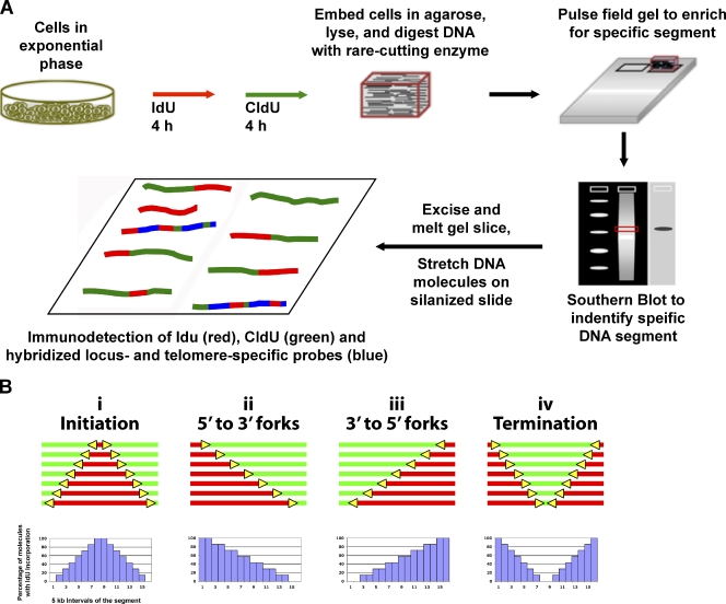
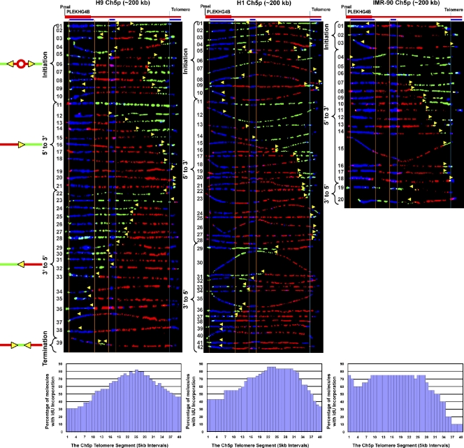
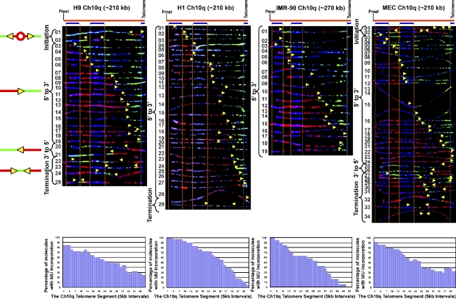
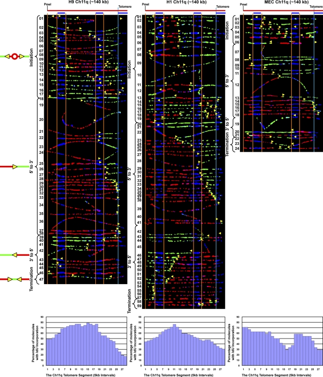
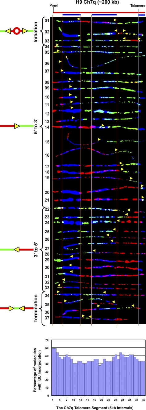
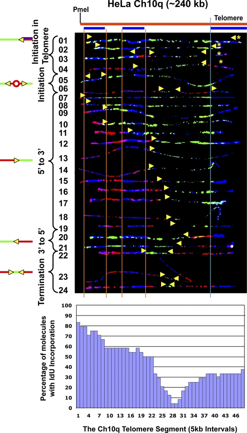
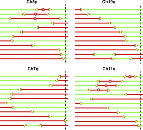
Telomeric and adjacent subtelomeric heterochromatin pose significant challenges to the DNA replication machinery. Little is known about how replication progresses through these regions in human cells. Using single molecule analysis of replicated DNA (SMARD), we delineate the replication programs-i.e., origin distribution, termination site location, and fork rate and direction-of specific telomeres/subtelomeres of individual human chromosomes in two embryonic stem (ES) cell lines and two primary somatic cell types. We observe that replication can initiate within human telomere repeats but was most frequently accomplished by replisomes originating in the subtelomere. No major delay or pausing in fork progression was detected that might lead to telomere/subtelomere fragility. In addition, telomeres from different chromosomes from the same cell type displayed chromosome-specific replication programs rather than a universal program. Importantly, although there was some variation in the replication program of the same telomere in different cell types, the basic features of the program of a specific chromosome end appear to be conserved.


Figures

Figure 1.
Figure 1.
SMARD. (A) Exponentially growing cells were labeled with the halogenated nucleosides IdU for 4 h followed by CldU for 4 h. Genomic DNA was then isolated. To avoid the breakage of large molecules, the cells were embedded in agarose. We used rare cutting restriction endonucleases to obtain unique large molecules of genomic DNA, which were separated by pulse field gel electrophoresis to enrich by size for the particular subtelomere segments of interest. Southern blotting was performed to identify the target segment within the gel, which was excised. The gel slice containing the segment of interest was melted and the enriched DNA in the melted gel solution was stretched on silanized glass slides. Fluorescent antibodies were used to identify regions where IdU (red) or CldU (green) were incorporated into the DNA. Immunodetected biotinylated FISH probes (blue) were used to identify the target molecules and to align the images of individual molecules to produce a composite profile of replication. (B) Specific replication events are identified by characteristic patterns in the aligned molecule images. DNA molecules with increasing red stain from one end indicate replication forks progressing in a single direction through the aligned region (ii and iii). Initiation events are indicated by a red tract flanked on both sides by green (i), whereas a green tract flanked on both sides by red (iv) indicates a termination event. Molecule alignments can be graphically depicted as histograms of the percentage of IdU incorporation along each 5 kb of a segment. Specific replication events produce characteristic features in these replication profiles. Positions of the centers of initiation zones are indicated by peaks (i), whereas termination events are indicated by valleys (iv). Replication progressing through the segment primarily in one direction (5′ to 3′ or 3′ to 5′) from an external origin is observed as a progressive decrease in the percentage of IdU incorporation from one end to the other across the segment (x axis; ii and iii).
Figure 2.
Figure 2.
Initiation events occur throughout the subtelomere in the human 5p segment in three cell lines, and replication forks progress through the segment in both directions. SMARD analysis of three different cell lines indicated that the 5p telomere is replicated primarily by forks progressing from the subtelomere to the telomere. Alignments of replicated molecules fully labeled with both IdU (red) and CldU (green) are shown. A map of the 5p locus is depicted above each alignment, with the positions of the FISH probes (blue bars below) used for identifying and orienting the molecules indicated. Vertical orange lines indicate the positions of the ends of the subtelomeric FISH signals used to align the molecules. The boundary between the subtelomere and telomere is delineated by a vertical blue line. Yellow arrows mark sites of transition from IdU incorporation to CldU incorporation and indicate the direction of fork progression at the moment of transition during the replication of the molecule. Replication profiles, histograms of the percentage of molecules containing IdU per 5-kb interval along the segment, are shown below each alignment. Initiation events (red tracts surrounded by green) occur at multiple locations. The origins appear to be clustered around a 40–90-kb region centered 65–115-kb from the telomere, seen as a peak in the replication profiles. Aside from a leftward broadening of the initiation zone in IMR-90 cells, there was not much variation in the basic features of the replication program of the 5p telomere in the two ES cell lines (H1 and H9) and the primary fibroblasts (IMR-90).
Figure 3.
Figure 3.
Replication forks progress toward the telomere from origins in the subtelomere of chromosome 10q in three cell types. SMARD analysis of the 10q telomere segment in ES lines H9 and H1, primary lung fibroblast, IMR-90 cells, and primary microvascular endothelial MECs indicated that the four cell lines replicate this segment using very similar programs. In these lines, the 10q telomere is replicated primarily by forks progressing from the subtelomere to the telomere. This is reflected in the replication profile histograms, which show a progressive decrease in the percentage of IdU incorporation from 5′ to 3′ across the segment, which is indicative of replication progressing through the segment primarily in one direction from an external origin. These distal subtelomeric origins are located predominantly >200 kb from the telomere in all four lines. A region of mixed staining (blue-green or blue-red) is seen in the right half of many molecules resulting from nonspecific hybridization of the FISH probes. The yellow arrows indicate the direction of replication fork progression, and the vertical orange and blue lines demarcate the boundaries of sequences where fish probes bind, as described in the legend to Fig. 2.
Figure 4.
Figure 4.
Initiation events occur throughout the human 11q segment and forks move across the segment in both directions in three cell lines.SMARD analysis of the Ch11q telomere segment in human ES H9, H1, and IMR-90 fibroblast cells revealed similar program features. Frequent initiation events occurred throughout the full 140-kb length of the segment. Preferred sites of initiation, seen as peaks in the replication profile histograms, could be detected. In all three lines, the 11q telomere is replicated primarily by forks progressing from the subtelomere to the telomere. The yellow arrows indicate the direction of replication fork progression, and the vertical orange and blue lines demarcate the boundaries of sequences where fish probes bind, as described in the legend to Fig. 2.
Figure 5.
Figure 5.
Initiation sites were rarely detected in the subtelomere of Ch 7q. SMARD analysis of the Ch 7 telomere/subtelomere segment of human ES H9 cells is shown. Few initiation or termination events were detected in the molecules examined. The lack of distinct peaks or valleys in the replication profile histogram indicates the absence of preferred initiation or termination sites. Similar numbers of replication forks proceed in the telomere-to-subtelomere (3′-to-5′) and subtelomere-to-telomere (5′-to-3′) directions. Many of the 5′-to-3′ forks appear to initiate from origins at least 150 kb from the telomere. The yellow arrows indicate the direction of replication fork progression, and the vertical orange and blue lines demarcate the boundaries of sequences where fish probes bind, as described in the legend to Fig. 2.
Figure 6.
Figure 6.
Replication forks initiate within the telomere repeats in chromosome 10q in a HeLa cell line with long telomeres. SMARD analysis of the Ch10q telomere segment in HeLa 1.3, a line with a mean telomere length of 23 kb, more than twice as long as in most human cells (Takai et al., 2010). Based on the length of the signals for the FISH probes, we estimate that the mean length of the telomeres in Ch10q is ∼40 kb. Telomeric initiation events are detected in molecules containing terminations where the red tract of the 3′-to-5′ fork does not extend out of the telomere or extends only a few kilobases out from the telomere (molecules 1–4). The centers of telomeric initiation events are indicated by yellow asterisks. A termination zone, seen as a valley in the replication profile histogram centered ∼60 kb from the telomere/subtelomere border, indicates the frequent use of two origins separated by at least 120 kb, which fire at similar times to replicate the segment. The yellow arrows indicate the direction of replication fork progression, and the vertical orange and blue lines demarcate the boundaries of sequences where fish probes bind, as described in the legend to Fig. 2.
Figure 7.
Figure 7.
Schematic representation of the replication program of four different telomere/subtelomere segments of the ES and primary somatic cells studied here. This diagrammatic summary of representative molecules indicates that human telomeres do not exhibit a universal replication program. The dotted vertical line indicates the boundary between the telomere and subtelomere.

Similar articles

Cited by

References

    1. Aladjem M.I. 2007. Replication in context: dynamic regulation of DNA replication patterns in metazoans. Nat. Rev. Genet. 8:588–600 10.1038/nrg2143 - DOI - PubMed
    1. Anand R.P., Shah K.A., Niu H., Sung P., Mirkin S.M., Freudenreich C.H. 2012. Overcoming natural replication barriers: differential helicase requirements. Nucleic Acids Res. 40:1091–1105 10.1093/nar/gkr836 - DOI - PMC - PubMed
    1. Arnoult N., Schluth-Bolard C., Letessier A., Drascovic I., Bouarich-Bourimi R., Campisi J., Kim S.H., Boussouar A., Ottaviani A., Magdinier F., et al. 2010. Replication timing of human telomeres is chromosome arm-specific, influenced by subtelomeric structures and connected to nuclear localization. PLoS Genet. 6:e1000920 10.1371/journal.pgen.1000920 - DOI - PMC - PubMed
    1. Atanasiu C., Deng Z., Wiedmer A., Norseen J., Lieberman P.M. 2006. ORC binding to TRF2 stimulates OriP replication. EMBO Rep. 7:716–721 10.1038/sj.embor.7400730 - DOI - PMC - PubMed
    1. Azzalin C.M., Reichenbach P., Khoriauli L., Giulotto E., Lingner J. 2007. Telomeric repeat containing RNA and RNA surveillance factors at mammalian chromosome ends. Science. 318:798–801 10.1126/science.1147182 - DOI - PubMed

Publication types