. 2012 Apr 16;197(2):301-12.
doi: 10.1083/jcb.201109132.

Munc13-4 reconstitutes calcium-dependent SNARE-mediated membrane fusion

Affiliations

Munc13-4 reconstitutes calcium-dependent SNARE-mediated membrane fusion

Kristin L Boswell et al. J Cell Biol. .

Abstract

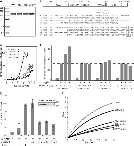
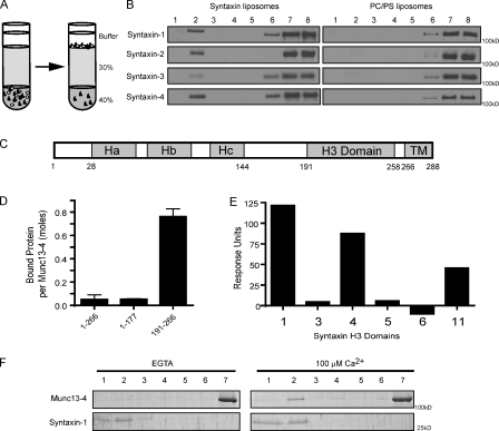
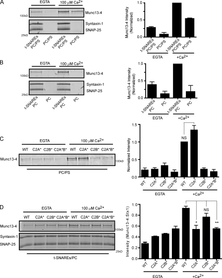
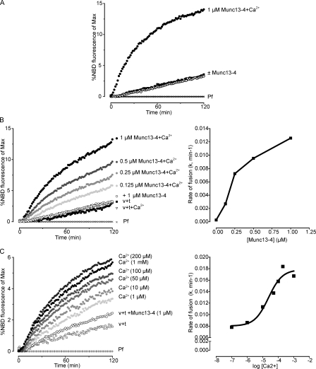
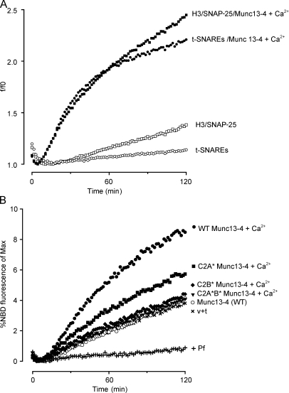
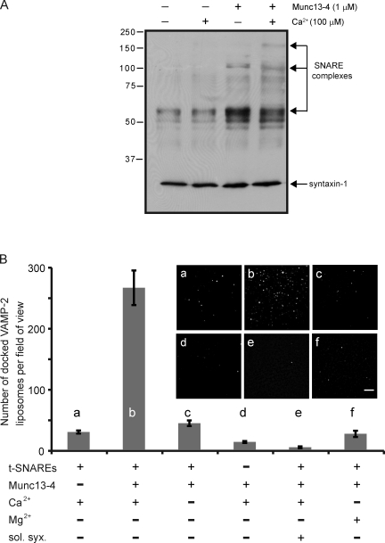
Munc13-4 is a widely expressed member of the CAPS/Munc13 protein family proposed to function in priming secretory granules for exocytosis. Munc13-4 contains N- and C-terminal C2 domains (C2A and C2B) predicted to bind Ca(2+), but Ca(2+)-dependent regulation of Munc13-4 activity has not been described. The C2 domains bracket a predicted SNARE-binding domain, but whether Munc13-4 interacts with SNARE proteins is unknown. We report that Munc13-4 bound Ca(2+) and restored Ca(2+)-dependent granule exocytosis to permeable cells (platelets, mast, and neuroendocrine cells) dependent on putative Ca(2+)-binding residues in C2A and C2B. Munc13-4 exhibited Ca(2+)-stimulated SNARE interactions dependent on C2A and Ca(2+)-dependent membrane binding dependent on C2B. In an apparent coupling of membrane and SNARE binding, Munc13-4 stimulated SNARE-dependent liposome fusion dependent on putative Ca(2+)-binding residues in both C2A and C2B domains. Munc13-4 is the first priming factor shown to promote Ca(2+)-dependent SNARE complex formation and SNARE-mediated liposome fusion. These properties of Munc13-4 suggest its function as a Ca(2+) sensor at rate-limiting priming steps in granule exocytosis.


Figures

Figure 1.
Figure 1.
Munc13-4 reconstitutes Ca2+-dependent granule exocytosis in multiple cell types. (A) Coomassie-stained SDS gels of wild-type (WT) and C2A*, C2B*, and C2A*B* Munc13-4 proteins. A minor degradation fragment was present in some preparations. Molecular mass is indicated in kilodaltons. (B) A schematic representation of the Munc13-4 sequence showing central DUF1041 and MHD1 homology domains present in all CAPS/Munc13 proteins and the MHD2 homology domain conserved in Munc13 proteins. A pileup of sequences from the β1-β2 regions of the C2A and C2B domains of hMunc13-4 and hSynaptotagmin-1 is shown with the conserved aspartate residues in boxes. Positions of the asparagine substitutions in C2A (D127N and D133N) and in C2B (D941N and D947N) are indicated. (C) Ca2+ binding to Munc13-4 measured by changes of intrinsic tryptophan fluorescence. 2 µM wild-type, C2A*, C2B*, and C2A*B* Munc13-4 proteins were excited at 280 nm, and emission spectra were collected from 290–390 nm. Normalized plots of the percentage change in fluorescence intensity are shown. These indicated an ∼5 µM [Ca2+]1/2 for wild-type Munc13-4, whereas [Ca2+]1/2 values for C2A*, C2B*, and C2A*B* Munc13-4 proteins exceeded 300 µM. Mean ± SD values are shown (n = 3). (D) [3H]serotonin release (as a percentage of the total) from preloaded permeable platelets is shown in the absence or presence of 10 µM Ca2+ at the indicated concentrations of added wild-type or mutant Munc13-4 proteins. The dotted line shows release in the absence of added Munc13-4. Results are representative of three similar experiments. (E) Release of ANF-GFP (as a percentage of the total) from mechanically permeabilized RBL-2H3 cells is shown for incubations without or with 10 µM Ca2+, 0.5 mg/ml cytosol, or 10 nM Munc13-4 or C2 domain mutants. Munc13-4 addition without Ca2+ was not stimulatory. Results shown are the mean ± SD for triplicate determinations in a single experiment. The inset shows 120-kD Munc13-4 extracted from intact or permeabilized washed RBL-2H3 cells analyzed by SDS-PAGE and immunoblotting. (F) Release of norepinephrine from mechanically permeabilized PC12 cells detected by voltammetry. Incubations at 35°C were conducted in the presence of 20 nM CAPS, 100 nM Munc13-4, or Munc13-4 mutants, as indicated. No release occurs until Ca2+ (to ∼1 µM) was injected (at time 0). Each curve represents a single sample. Results are representative of three similar experiments.
Figure 2.
Figure 2.
Munc13-4 binds to the H3 region of syntaxins. (A) Bound and free Munc13-4 were determined by buoyant density flotation of liposomes. (B) 1 µM Munc13-4 was incubated with PC/PS liposomes containing syntaxin-1, -2, -3, or -4 (left) or protein-free PC/PS liposomes (right) for 30 min at room temperature. Bound (fraction 2) and free (fractions 6–8) liposomes were separated on Accudenz gradients and analyzed for Munc13-4 by immunoblotting. Results are representative of two similar experiments. Densitometry with ImageQuant software indicated that bound/free liposomes corresponded to 0.11, 0.06, 0.03, and 0.17 for syntaxin-1, -2, -3, and -4, respectively. (C) Syntaxin-1 contains Habc(28–144), SNARE motif/H3(191–258), and transmembrane(266–288) (TM)domains. (D) Surface plasmon resonance experiments of syntaxin-1 fragments (1–266, 1–177, and 191–266) binding to Munc13-4. Specific binding to Munc13-4 was determined by subtracting background binding to the control MBP. Mean values ± SEM are shown (n = 3). (E) Surface plasmon resonance experiments of the H3 domains of syntaxin-1, -3, -4, -5, -6, and -11. Specific binding to Munc13-4 was determined by subtracting background binding to the control MBP. Results are representative of two similar experiments. (F) 1 µM Munc13-4 was incubated with syntaxin-1–containing liposomes composed of PC/PS/PIP2 (85:12:3) in the presence of EGTA or 100 µM free Ca2+. Accudenz gradient fractions were analyzed by SDS-PAGE and stained with SYPRO ruby.
Figure 3.
Figure 3.
Ca2+-regulated Munc13-4 binding to t-SNAREs and PS requires C2A and C2B domains, respectively. (A–D) All binding experiments were conducted by incubating 1 µM Munc13-4 with the indicated liposomes at room temperature for 30 min with EGTA or Ca2+ followed by separation of bound and free protein by buoyant density centrifugation conducted in the absence or presence of Ca2+. Bound fractions were analyzed by SDS-PAGE, stained with SYPRO ruby, and quantified by densitometry. Munc13-4 migrated between 100- and 150-kD standards. Syntaxin-1 and SNAP-25 migrated between 25- and 37-kD standards. The extent of binding (right graphs) is shown as means ± SEM for n = 3. (A) Munc13-4 binding to t-SNARE–containing or protein-free PC/PS liposomes in the presence of EGTA or 100 µM Ca2+. (B) Munc13-4 binding to t-SNARE–containing or protein-free PC liposomes in the presence of EGTA or 100 µM Ca2+. (C) Binding of wild-type (WT), D127N/D133N, D941N/D947N, and D127N/D133N/D941N/D947N Munc13-4 proteins (termed WT, C2A*, C2B*, and C2A*B* Munc13-4, respectively) to PC/PS liposomes in the presence of EGTA or 100 µM Ca2+. Binding of WT and C2A* Munc13-4 to PC/PS liposomes in the presence of Ca2+ was not significantly different. (D) Binding of wild-type, C2A*, C2B*, and C2A*B* Munc13-4 proteins to t-SNARE–containing PC liposomes. Munc13-4 binding was normalized to syntaxin-1 (Stx1) content of liposomes. Ca2+-stimulated binding of C2A* and C2A*B* Munc13-4 proteins was significantly reduced compared with wild type (*, P < 0.05; **, P < 0.002), whereas binding by C2B* Munc13-4 was not significantly different based on an unpaired t test.
Figure 4.
Figure 4.
Munc13-4 promotes liposome fusion in a calcium-dependent manner. (A) VAMP-2–containing PC/PS donor liposomes (with NBD-PE and Rh-PE) and t-SNARE–containing PC/PS acceptor liposomes were incubated with 1 µM Munc13-4 in the presence of EGTA or 400 µM free calcium (+Ca2+), as indicated. Control reactions with protein-free (Pf) liposomes replacing t-SNARE liposomes were incubated in parallel for background subtraction. Fusion was detected as increased NBD-PE fluorescence relative to maximal dequenched values. (B) Incubations similar to those of A were conducted at 100 µM Ca2+ at the indicated Munc13-4 concentrations (left), and initial fusion rates were determined (right). v + t, reactions with v-SNARE–containing liposomes plus t-SNARE–containing liposomes. (C) Incubations similar to those of A were conducted with 1 µM Munc13-4 at the indicated free Ca2+ concentrations (left), and initial fusion rates were determined (right). The EC50 for Ca2+ was calculated to be 23 ± 4 µM. In all graphs, the curves represent single samples. Results shown are representative of two to four similar experiments.
Figure 5.
Figure 5.
Munc13-4 utilizes N-terminally truncated syntaxin-1 and requires Ca2+-binding C2 domains in SNARE-dependent liposome fusion. (A) Incubations were conducted in the absence or presence of 1 µM Munc13-4 and 400 µM Ca2+, as indicated with DiI-containing VAMP-2 donor liposomes and DiD-containing acceptor liposomes that contained either syntaxin-1(191–288) with SNAP-25 (H3/SNAP-25) or full-length syntaxin-1(1–288) with SNAP-25 (t-SNARE). Fusion was detected as increased DiD fluorescence expressed relative to the lowest fluorescence as a ratio (f/fo). Curves show individual samples. Results are representative of three similar experiments. (B) 1 µM wild-type (WT), C2A*, C2B*, or C2A*B* Munc13-4 proteins were incubated with donor v-SNARE liposomes (containing NBD-PE and Rh-PE) and acceptor t-SNARE liposomes, as in Fig. 4 A, with 100 µM Ca2+. Fusion was detected as increased NBD-PE fluorescence relative to maximal dequenched values. Curves show individual samples. Results are representative of two similar experiments. v + t, reactions with v-SNARE–containing liposomes plus t-SNARE–containing liposomes.
Figure 6.
Figure 6.
Munc13-4 stimulation of trans-SNARE complex formation is Ca2+ dependent. (A) Munc13-4 stimulates the formation of SDS-resistant SNARE complexes. Incubations with VAMP-2– and syntaxin-1/SNAP-25–containing liposomes were conducted on ice for 30 min with 100 µM Ca2+ and 1 µM Munc13-4, as indicated. Reactions terminated in SDS sample buffer without boiling were analyzed by immunoblotting with monoclonal HPC-1 syntaxin-1 antibody. The arrows indicate the positions of ∼70-kD, ∼120-kD, and ∼165-kD SNARE complexes. Similar experiments indicated that VAMP-2 and SNAP-25 comigrated with these complexes. See James et al. (2009) for characterization of SNARE protein complexes. (B) Munc13-4 stimulates the docking of VAMP-2 liposomes onto t-SNARE–containing supported bilayer membranes. VAMP-2 liposomes with Rh-PE were incubated with t-SNARE–containing supported bilayers, and the number of liposomes stably docked for ≥2 min was monitored by total internal reflection fluorescence microscopy. Bar, 5 µm. Incubations contained 1 µM Munc13-4, 200 µM Ca2+, or 200 µM Mg2+, as indicated. Munc13-4 promoted liposome docking in the presence of Ca2+ (b) but not in its absence (c) or with Mg2+ (f), nor was liposome docking promoted by Ca2+ alone (a). t-SNAREs were required for Munc13-4–promoted docking (d), whereas preincubations of t-SNARE bilayers with 8 µM soluble syntaxin(1–266) (sol. syx.) for 30 min (e) inhibited Munc13-4–promoted docking. Mean values ± SD (n = 3) are shown.

References

    1. Arneson L.N., Brickshawana A., Segovis C.M., Schoon R.A., Dick C.J., Leibson P.J. 2007. Cutting edge: Syntaxin 11 regulates lymphocyte-mediated secretion and cytotoxicity. J. Immunol. 179:3397–3401 - PubMed
    1. Augustin I., Rosenmund C., Südhof T.C., Brose N. 1999. Munc13-1 is essential for fusion competence of glutamatergic synaptic vesicles. Nature. 400:457–461 10.1038/22768 - DOI - PubMed
    1. Banerjee A., Kowalchyk J.A., DasGupta B.R., Martin T.F. 1996. SNAP-25 is required for a late postdocking step in Ca2+-dependent exocytosis. J. Biol. Chem. 271:20227–20230 10.1074/jbc.271.45.28294 - DOI - PubMed
    1. Bryceson Y.T., Rudd E., Zheng C., Edner J., Ma D., Wood S.M., Bechensteen A.G., Boelens J.J., Celkan T., Farah R.A., et al. 2007. Defective cytotoxic lymphocyte degranulation in syntaxin-11 deficient familial hemophagocytic lymphohistiocytosis 4 (FHL4) patients. Blood. 110:1906–1915 10.1182/blood-2007-02-074468 - DOI - PMC - PubMed
    1. Chen D., Bernstein A.M., Lemons P.P., Whiteheart S.W. 2000. Molecular mechanisms of platelet exocytosis: Role of SNAP-23 and syntaxin 2 in dense core granule release. Blood. 95:921–929 - PubMed

Publication types

MeSH terms