. 2012 Aug;27(8):1746-56.
doi: 10.1002/jbmr.1637.

An Acvr1 R206H knock-in mouse has fibrodysplasia ossificans progressiva

Affiliations

An Acvr1 R206H knock-in mouse has fibrodysplasia ossificans progressiva

Salin A Chakkalakal et al. J Bone Miner Res. 2012 Aug.

Abstract

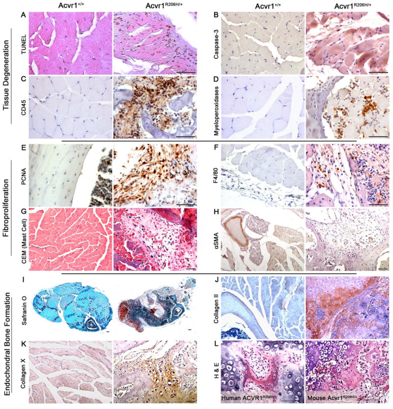
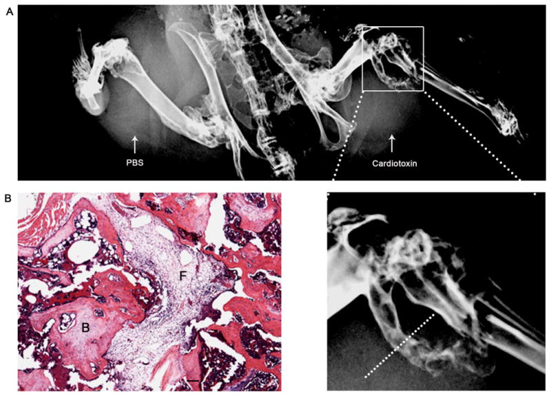
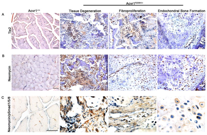
Fibrodysplasia ossificans progressiva (FOP; MIM #135100) is a debilitating genetic disorder of dysregulated cellular differentiation characterized by malformation of the great toes during embryonic skeletal development and by progressive heterotopic endochondral ossification postnatally. Patients with these classic clinical features of FOP have the identical heterozygous single nucleotide substitution (c.617G > A; R206H) in the gene encoding ACVR1/ALK2, a bone morphogenetic protein (BMP) type I receptor. Gene targeting was used to develop an Acvr1 knock-in model for FOP (Acvr1(R206H/+)). Radiographic analysis of Acvr1(R206H/+) chimeric mice revealed that this mutation induced malformed first digits in the hind limbs and postnatal extraskeletal bone formation, recapitulating the human disease. Histological analysis of murine lesions showed inflammatory infiltration and apoptosis of skeletal muscle followed by robust formation of heterotopic bone through an endochondral pathway, identical to that seen in patients. Progenitor cells of a Tie2(+) lineage participated in each stage of endochondral osteogenesis. We further determined that both wild-type (WT) and mutant cells are present within the ectopic bone tissue, an unexpected finding that indicates that although the mutation is necessary to induce the bone formation process, the mutation is not required for progenitor cell contribution to bone and cartilage. This unique knock-in mouse model provides novel insight into the genetic regulation of heterotopic ossification and establishes the first direct in vivo evidence that the R206H mutation in ACVR1 causes FOP.


Conflict of interest statement

Disclosure Statement: ADAM received grant support from Hologic Inc. (Bedford, MA). All the other authors state that they have no conflicts of interest.

Figures

Figure 1
Figure 1. Generation of Acvr1R206H/+ knock-in mouse model for FOP
(A) Targeting strategy. Schematic representation of the wild-type Acvr1 locus, the targeting construct with the R206H substitution in exon 5, and the inserted mutant allele following homologous recombination. Exons (black boxes), BglII restriction sites, probes for Southern detection, and the neomycin selection cassette flanked by FLP sites are shown. (B) Homologous recombination detection by Southern analysis of BglII-cleaved genomic DNA from ES cell clones. Homologous recombination was indicated by detection of mutant (14.4 kb) and wild-type (12.4 kb) alleles (example circled). (C) DNA sequence analysis of heterozygous Acvr1 alleles in recombined ES cells (right panel); wild-type sequence (left panel). Position c.617 is indicated by arrows. [A color version of this figure is available as online Supplemental Information.]
Figure 2
Figure 2. Acvr1R206H/+mice display classic FOP phenotypes
(A) Characteristic great toe malformation in FOP. Gross anatomical (left panel) and X-ray (right panel) images from a patient with the classic Acvr1R206H/+ FOP mutation. (B) Acvr1R206H/+ chimeric mice (right panels; representative 3-month old mouse) displayed malformation of the first digits of the hind-limbs (circled) at birth. X-ray analyses revealed shortened or absent proximal and distal phalanges which are comparable to the malformations observed in FOP patients. (C-F) μCT shows extraskeletal bone formation and skeletal malformations associated with FOP (a representative Acvr1R206H/+ mouse at 8 weeks old). Whole body image (C) with arrows to indicate extra-skeletal bone formation is shown. Fusion of cervical vertebrae (C3, C4, C5) (D), costovertebral malformations and fusion of vertebrae (asterisks) (E), and osteochondromas (arrows) (F) are observed in the mouse and FOP patients. [A color version of this figure is available as online Supplemental Information.]
Figure 3
Figure 3. Histological analysis of cellular events during heterotopic bone formation in Acvr1R206H/+ knock-in chimeric mice
Skeletal muscle tissue sections from chimeric Acvr1R206H/+ mice (at 2 months of age) are shown in comparison to control tissues from wild-type mice. (A-D) Initiation of heterotopic bone formation is associated with degeneration of soft connective tissues. (A) TUNEL staining revealed DNA fragmentation (brown staining) that is characteristic of apoptosis. (B) Apoptosis was confirmed by the positive staining for activated caspase-3; myocentric nuclei (arrow) in degenerating myofibers are observed. (C) Immunohistochemical staining for CD45 (lymphocyte marker) supports immune system cell participation in early stages of lesion formation. (D) Immunohistochemical staining for myeloperoxidase, a marker for neutrophils, was detected around degenerating myofibers. (E-H) A fibroproliferative stage follows tissue degeneration. (E) Regions containing fibroblasts stained positively for PCNA indicating robust proliferation. In control tissues, abundant PCNA positive cells were located within the bone marrow cavity. (F) Immunostaining with F4/80 antibodies detected monocytes/macrophages in the fibroproliferative areas; these cells were also observed in degenerating tissues. (G) Activated granular mast cells (blue CEM staining) were abundant in fibroproliferative and degenerative stages. Occasional non-activated agranular mast cells were detected in control sections. (H) Immunostaining for alpha smooth muscle actin (αSMA) indicated angiogenesis and new blood vessel formation in fibroproliferative areas. (I-L) Heterotopic endochondral bone formation in Acvr1R206H/+ mice. Low power images of a cross section through heterotopic lesion were stained with safranin O (I) to show chondrocytes and cartilage. (J) Cartilage is positive for collagen II and (K) hypertrophic chondrocytes were detected by collagen X. (L) High power images (H&E) of late stage lesions from an FOP patient (left panel) and mouse (right panel) show mature chondrocytes and bone formation. Scale bar: 50 μm for all images (except panel I bar = 200 μm).
Figure 4
Figure 4. Skeletal muscle injury induces heterotopic ossification in Acvr1R206H/+ mice
Skeletal muscle was injured by cardiotoxin injection into the hind limbs of Acvr1R206H/+ mice and examined 6 weeks after injury. (A) X-ray images revealed severe heterotopic bone formation in response to cardiotoxin but not PBS. (B) Histological analysis (H&E) of a limb cross section showed the range of stages of lesion formation including newly formed heterotopic bone. Scale bar: 50 μm.
Figure 5
Figure 5. Cells participating in heterotopic lesion formation
Skeletal muscle and areas of heterotopic ossification, similar to Figure 3, are shown. (A) Tie2-positive lineage cells contribute to heterotopic lesion formation. Cells positive for Tie2, an endothelial cell marker, are present throughout lesion formation as shown in tissues from the degeneration, fibroproliferative, and endochondral bone formation stages in Acvr1R206H/+ mice. Tie2+ cells were undetected in skeletal muscle from wild-type mice. Scale bar: 50 μm. (B) The Acvr1R206H/+ mutation is not required in cells that differentiate into ectopic cartilage and bone. In chimeric Acvr1R206H/+ mice, mutant cells are distinguished from wild-type by neomycin phosphotransferase II. Representative images from Acvr1R206H/+ mice show both neomycin positive and neomycin negative (wild-type) cells in the degenerative, fibroproliferative, and chondrogenesis stages, indicating the participation of both mutant and wild-type cells in the progression of heterotopic ossification lesion formation. Tissues from wild-type mice (Acvr1+/+; right panel) showed no staining for neomycin phosphotransferase II. Scale bar: 50 μm. (C) Correlation of neo expressing cells (Acvr1R206H/+) with BMP signaling. Lesional areas of tissue degradation, fibroproliferative, and endochondral ossification stages were detected for neomycin (brown, cytoplasmic staining) and pSmad1/5/8 (blue, nuclear staining) by double immunohistochemistry. Double positive cells are observed in all stages of lesion development and were the most abundant cells present (also see Supplemental Figure 3). Scale bar: 50 μm.

References

    1. Pignolo RJ, Foley KL. Nonhereditary Heterotopic Ossification: Implications for Injury, Arthropathy, and Aging. Clin Rev Bone Min Metab. 2005;3(3-4):261–6.
    1. McCarthy EF, Sundaram M. Heterotopic ossification: a review. Skeletal Radiol. 2005;34(10):609–19. - PubMed
    1. Forsberg JA, Pepek JM, Wagner S, et al. Heterotopic ossification in high-energy wartime extremity injuries: prevalence and risk factors. J Bone Joint Surg Am. 2009;91(5):1084–91. - PubMed
    1. Davis TA, O'Brien FP, Anam K, Grijalva S, Potter BK, Elster EA. Heterotopic ossification in complex orthopaedic combat wounds: quantification and characterization of osteogenic precursor cell activity in traumatized muscle. J Bone Joint Surg Am. 2011;93(12):1122–31. - PubMed
    1. Potter BK, Forsberg JA, Davis TA, et al. Heterotopic ossification following combat-related trauma. J Bone Joint Surg Am. 2010;92(2):74–89. - PubMed

Publication types

MeSH terms