. 2012 Jul;26(7):2986-99.
doi: 10.1096/fj.12-204495. Epub 2012 Apr 16.

Upregulation of proteasome activity in muscle RING finger 1-null mice following denervation

Affiliations

Upregulation of proteasome activity in muscle RING finger 1-null mice following denervation

Aldrin V Gomes et al. FASEB J. 2012 Jul.

Abstract

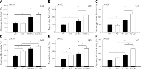
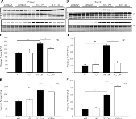
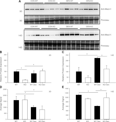
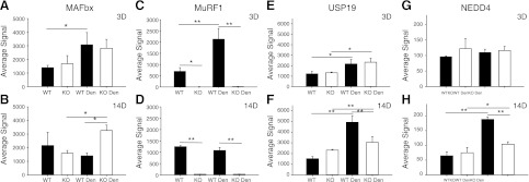
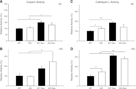
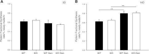
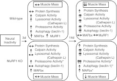
Deletion of muscle RING finger 1 (MuRF1), an E3 ubiquitin ligase, leads to sparing of muscle mass following denervation. The purpose of this study was to test the hypothesis that muscle sparing in mice with a deletion of MuRF1 is due to the selective inhibition of the ubiquitin proteasome system. Activities of the 20S and 26S proteasomes, calpain and cathepsin L, were measured in the triceps surae muscles of wild-type (WT) and MuRF1-knockout (KO) mice at 3 and 14 d following denervation. In addition, fractional protein synthesis rates and differential gene expression were measured in WT and KO muscle. The major finding was that 20S and 26S proteasome activities were significantly elevated (1.5- to 2.5-fold) after 14 d of denervation in both WT and KO mice relative to control, but interestingly, the activities of both the 20S and 26S proteasome were significantly higher in KO than WT mice. Further, mRNA expression of MAFbx was elevated after 14 d of denervation in KO, but not WT, mice. These data challenge the conventional dogma that MuRF1 is controlling the degradation of only contractile proteins and suggest a role for MuRF1 in the global control of the ubiquitin proteasome system and protein turnover.


Figures

Figure 1.
Figure 1.
Muscle sparing in MuRF1-KO but not MAFbx-KO mice. A) Representative muscle cross section from the TA muscle of WT, MuRF1-KO, and MAFbx-KO mice taken after 28 d of denervation. Muscle cross sections were stained with hematoxylin and eosin and show muscle fiber degeneration (arrows), primarily in the MAFbx-KO mice. B) Muscle mass, expressed as relative wet weight (denervated/control), is plotted for the TS complex (solid bars) and TA (open bars) muscles following 28 d of denervation. Data are expressed as means ± sd of muscles from WT (n=10), MuRF1-KO (n=7), and MAFbx-KO (n=7) mice. C) Muscle mass, expressed as relative wet weight (denervated/control), is plotted for the TS complex following 3 d (solid bars) and 14 d (open bars) of denervation. Data are means ± sd of muscles from WT (n=5–7/time point) and MuRF1-KO (n=7–8/time point) mice. *P < 0.05 vs. WT denervated muscle.
Figure 2.
Figure 2.
20S and 26S proteasome proteolytic activities in WT and MuRF1-KO mice after 3 d of denervation. ATP-dependent (26S; A–C) and ATP-independent (20S; D–F) activities in the TS muscles of WT (solid bars) and MuRF1-KO (open bars) mice following 3 d of denervation. Individual subunit activities are shown for β1, caspase-like activity (A, D); β2, trypsin-like activity (B, E); and β5, chymotrypsin-like activity (C, F). Data are expressed as means ± sd of muscles (n=4/group). *P < 0.05.
Figure 3.
Figure 3.
20S and 26S proteasome proteolytic activities in WT and MuRF1-KO mice after 14 d of denervation. ATP-dependent (26S; A–C) and ATP-independent (20S; D–F) activities in the TS muscles of WT (solid bars) and MuRF1-KO (open bars) mice following 14 d of denervation. Individual subunit activities are shown for β1, caspase-like activity (A, D); β2, trypsin-like activity (B, E); and β5, chymotrypsin-like activity (C, F). Data are means ± sd of muscles (n=4/group). *P < 0.05.
Figure 4.
Figure 4.
Protein expression of 19S and 20S proteasome subunits in control (con) and denervated (den) muscle. A, B) Western blots of lysates from the TS of WT (n=3–4) and MuRF1-KO (n=4) mice following 3 and 14 d of denervation. Immunoblots for PSMA6 (α1, 20S subunit; A, top panel) and Ponceau staining (A, bottom panel) and PSMC2 (Rpt1, 19S subunit; B, top panel) and Ponceau staining (B, bottom panel). C–F) Quantification of immunoblots from WT (solid bars) and MuRF1-KO (open bars) mice; data are expressed as means ± sd. Relative amounts (expressed as a percentage of WT con) of PSMA6 protein (C, E) or PSMC2 protein (D, F) in TS after 3 d (C, D) or 14 d (E, F) of denervation. *P < 0.05, **P < 0.001.
Figure 5.
Figure 5.
Protein and mRNA expression of the inducible proteasome subunit PSMB10 in control (con) and denervated (den) muscle. A) Western blots of lysates from the TS of WT (n=3–4) and MuRF1-KO (n=3–4) mice following 3 and 14 d of denervation. A) Immunoblot against anti-Mecl-1, antibody for the inducible proteasome subunit PSMB10 (top panel) and Ponceau staining (bottom panel). B, C) Quantification of immunoblots from WT (solid bars) and MuRF1-KO (open bars) mice; data are expressed as means ± sd. Relative amounts (expressed as a percentage of WT con) of PSMB10 protein in TS after 3 d (B) or 14 d (C) of denervation. D, E) Relative mRNA expression of the PSMB10 gene in the TS of WT (solid bars) and KO (open bars) mice after 3 d (D) or 14 d (E) of denervation. *P < 0.05.
Figure 6.
Figure 6.
Polyubiquitination levels in the TS muscle of WT and MuRF1-KO mice following denervation (den). A, B) Western blots of lysates from the TS of WT (n=3–4) and MuRF1-KO (n=4) mice following 3 (A) and 14 (B) d of denervation. Polyubiquitinated proteins were determined by immunoblotting with two antiubiquitin antibodies, FK1 and P4D1. C, D) Quantification of immunoblots from WT (solid bars) and MuRF1-KO (open bars) mice; data are expressed as means ± sd. Relative amounts (expressed as a percentage of control WT) of polyubiquitinated proteins in TS after 3 d (C) or 14 d (D) of denervation. *P < 0.05, **P < 0.001.
Figure 7.
Figure 7.
Differential mRNA expression in WT and MuRF1-KO mice following denervation (den). mRNA expression (average signal from Illumina microarray) of MAFbx (A, B), MuRF1 (C, D), USP19 (E, F) and NEDD4 (G, H) after 3 d (A, C, E, G) and 14 d (B, D, F, H) of denervation in WT (solid bars) and MuRF1-KO (open bars). Data are expressed as means ± sd from n = 3/group. *P < 0.05, **P < 0.001.
Figure 8.
Figure 8.
Calpain and cathepsin L activities in WT and MuRF1-KO mice following denervation (den). A, B) Calcium-dependent calpain activity in the TS muscle of WT (solid bars) and KO (open bars) mice after 3 (A) and 14 (B) d of denervation. C, D) Cathepsin L activity in the TS muscle of WT (solid bars) and KO (open bars) mice after 3 (C) and 14 (D) d of denervation. Data are expressed as means ± sd for n = 3 or 4 mice/group. Enzyme activity is expressed relative to the WT Con group. *P < 0.05, **P < 0.001.
Figure 9.
Figure 9.
Protein fractional synthesis rate (FSR) of the TS muscle of WT and MuRF1-KO mice following denervation (den). Protein FSR (% newly made/h) was measured in TS of WT (solid bars) and MuRF1-KO (open bars) mice after 3 (A) and 14 d of denervation (B). Data are means ± sd for n = 4–10. ** P < 0.001.
Figure 10.
Figure 10.
Summary of major findings. Diagrammatic representation of the major changes found in the TS complex of WT and MuRF1-KO mice following 3 and 14 d of denervation. Both proteolytic and synthesis pathways are significantly affected by denervation. Asterisks indicate novel and significant differences that were observed between WT and MuRF1-KO mice following denervation. The increased proteasome activity in MuRF1-KO mice following denervation was unexpected and runs contrary to current dogma. We propose that the increase in protein synthesis in MuRF1-KO mice is through different pathways than occurs in WT mice. Further studies are required to identify the specific protein targets of MuRF1. Note that the most significant differences between the WT and MuRF1-KO mice do not occur at 3 d, when MuRF1 mRNA expression in the WT is at a peak, but at 14 d, after MuRF1 mRNA expression has gone back to baseline.

References

    1. Medina R., Wing S. S., Goldberg A. L. (1995) Increase in levels of polyubiquitin and proteasome mRNA in skeletal muscle during starvation and denervation atrophy. Biochem. J. 307, 631–637 - PMC - PubMed
    1. Taillandier D., Aurousseau E., Meynial-Denis D., Bechet D., Ferrara M., Cottin P., Ducastaing A., Bigard X., Guezennec C. Y., Schmid H. P., Attaix D. (1996) Coordinate activation of lysosomal, Ca2+-activated and ATP-ubiquitin-dependent proteinases in the unweighted rat soleus muscle. Biochem. J. 316, 65–72 - PMC - PubMed
    1. Price S. R., Bailey J. L., Wang X., Jurkovitz C., England B. K., Ding X., Phillips L. S., Mitch W. E. (1996) Muscle wasting in insulinopenic rats results from activation of the ATP-dependent, ubiquitin–proteasome proteolytic pathway by a mechanism including gene transcription. J. Clin. Invest. 98, 1703–1708 - PMC - PubMed
    1. Temparis S., Asensi M., Taillandier D., Aurousseau E., Larbaud D., Obled A., Bechet D., Ferrara M., Estrela J. M., Attaix D. (1994) Increased ATP-ubiquitin-dependent proteolysis in skeletal muscles of tumor-bearing rats. Cancer Res. 54, 5568–5573 - PubMed
    1. Baracos V. E., DeVivo C., Hoyle D. H., Goldberg A. L. (1995) Activation of the ATP-ubiquitin-proteasome pathway in skeletal muscle of cachectic rats bearing a hepatoma. Am. J. Physiol. 268, E996–E1006 - PubMed

Publication types

MeSH terms

LinkOut - more resources