. 2012 Apr 15;26(8):803-15.
doi: 10.1101/gad.187641.112.

The ubiquitin ligase mLin41 temporally promotes neural progenitor cell maintenance through FGF signaling

Affiliations

The ubiquitin ligase mLin41 temporally promotes neural progenitor cell maintenance through FGF signaling

Jianfu Chen et al. Genes Dev. .

Erratum in

  • Genes Dev. 2012 Jun 15;26(12):1392

Abstract

How self-renewal versus differentiation of neural progenitor cells is temporally controlled during early development remains ill-defined. We show that mouse Lin41 (mLin41) is highly expressed in neural progenitor cells and its expression declines during neural differentiation. Loss of mLin41 function in mice causes reduced proliferation and premature differentiation of embryonic neural progenitor cells. mLin41 was recently implicated as the E3 ubiquitin ligase that mediates degradation of Argonaute 2 (AGO2), a key effector of the microRNA pathway. However, our mechanistic studies of neural progenitor cells indicate mLin41 is not required for AGO2 ubiquitination or stability. Instead, mLin41-deficient neural progenitors exhibit hyposensitivity for fibroblast growth factor (FGF) signaling. We show that mLin41 promotes FGF signaling by directly binding to and enhancing the stability of Shc SH2-binding protein 1 (SHCBP1) and that SHCBP1 is an important component of FGF signaling in neural progenitor cells. Thus, mLin41 acts as a temporal regulator to promote neural progenitor cell maintenance, not via the regulation of AGO2 stability, but through FGF signaling.


Figures

Figure 1.
Figure 1.
mLin41 expression declines during neural differentiation. (A) Western blot analyses of the protein expression of mLin41 and neural markers in cranial neuroepithelium at different embryonic stages as indicated (represented by “E” for days post-coitum). β-Actin serves as a loading control. (B,C) In situ hybridization analyses of mLin41 mRNA expression in sections of E9.5 and E12.5 embryos. Bars, 0.1 mM. (D–G) Whole-mount X-gal-stained embryos at E7.5 (D), E8.5 (E), E9.5 (F), and E10.5 (G). Red arrows indicate the absence of mLin41 expression in the heart at E9.5 and E10.5. Bars, 0.5 mM.
Figure 2.
Figure 2.
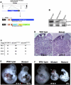
mLin41 is required for neural tube growth. (A) Genomic structure of mLin41 locus on mouse chromosome 9, and the gene trap vector pGT1lxf (BayGenomics) is inserted between exons 1 and 2. Wild-type mLin41 protein is ∼94 kDa, whereas the mutant protein arising from the fusion between the mLin41 N terminus (271 amino acids) and β-geo is ∼172 kDa. (B) Western blot analyses of mLin41 protein expression in cranial neuroepithelium of E9.5 wild-type, heterozygous, and mLin41lacZ/lacZ mutant embryos using an antibody that recognizes the C terminus of mLin41. β-Actin as loading control. (C) Number of wild-type (WT/WT), heterozygous (mLin41WT/lacZ), and mutant (mLin41lacZ/lacZ) embryos recovered alive from litters dissected at the indicated embryonic stages. (D) H&E staining of transverse sections from E10.5 wild-type and mutant neural tubes at comparable rostral–caudal levels. Black dots outline neural tubes, and small insets indicate the whole sections. Bar, 35 μm. (E) Ninety-two percent of E10.5 mutant embryos exhibit open neural tube (white arrow) and reduced neural tissue growth (red arrowhead) compared with littermate control embryos. Bar, 0.5 mM. (F) Eight percent of E10.5 mutants show dramatic growth retardation compared with littermate control embryos. Bar, 0.5 mM.
Figure 3.
Figure 3.
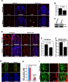
mLin41 deficiency leads to reduced cell proliferation in the neuroepithelium. (A) Confocal microscope images of sections from hindbrains and spinal cords of E9.5 wild-type and mLin41lacZ/lacZ mutant embryos. Anti-p-H3 antibody visualizes mitotic cells (red). White arrowheads indicate mislocalized p-H3-positive cells in the mutant neuroepithelium. Hoechst stains nuclei (blue). Bar, 200 μm. (B) Quantitation of p-H3-positive cells from A. Percentage of p-H3-positive cells was calculated as the percentage of p-H3-positive cells out of the total number of neuroepithelial cells, indicated by Hoechst staining, within the neural tube sections. Error bars indicate SEM of 12 sections from three independent experiments. P < 0.0001. (C) Western blot analyses of the expression level of p-H3 in E9.5 wild-type and mutant cranial neuroepithelium. β-Actin serves as a loading control. (D) Confocal microscope images of hindbrain and spinal cord sections from E9.5 wild-type and mutant embryos after 1 h of BrdU labeling. S-phase cells were visualized by anti-BrdU antibody (red); Hoechst stains nuclei (blue). Bar, 100 μm. (E,F) Quantitation of percentage of BrdU-positive cells per total number of neuroepithelial cells indicated by Hoechst staining in hindbrain and spinal cord sections. Error bars indicate SEM of 12 sections from three independent experiments. (G,I) Confocal microscope images of spinal cord sections from E9.5 wild-type and mutant embryos immunostained with anti-Ki67 antibody (green) to label cycling cells and anti-TuJ1 antibody (red) to label differentiated neurons. I is high magnification of the areas outlined by white boxes in G. White dots outline the neuroepithelial cells, and the white arrowhead indicates cells that coexpress Ki67 and TuJ1 in mutants. Bars: G, 200 μm; I, 50 μm. (H) Quantitative measurement of Ki67-positive cells or TuJ1-positive cells counted from each neural tube section of wild-type and mutant embryos. Error bars indicate SEM of nine sections from three independent experiments.
Figure 4.
Figure 4.
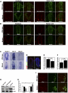
Loss of mLin41 results in premature differentiation of neural progenitor cells. (A,B) Confocal microscope images of sections from hindbrains and spinal cords of E9.5 wild-type and mLin41lacZ/lacZ mutant embryos. Bar, 200 μm. (A) Anti-Neurofilament antibody (red) marks differentiated neurons, and anti-Sox2 antibody (green) labels neural progenitor cells. (B) Anti-TuJ1 antibody (red) marks differentiated neurons, and anti-Nestin antibody (green) labels neural progenitor cells. Note that there are significantly more Neurofilament- and TuJ1-positive cells in the mutant neuroepithelium compared with littermate controls. (C) In situ hybridization on spinal cord sections of E9.5 wild-type and mutant embryos with probes for neural progenitor markers Notch1 and Hes-5 and differentiation marker Mash1. (D,I) Confocal microscope images of spinal cord sections from E9.5 wild-type and mutant embryos. Anti-Olig2 antibody (red) labels motor neuron precursors, and anti-Isl1/2 antibody (green) marks motor neurons. I is high magnification of the areas outlined by white boxes in D. White dots in I outline the neuroepithelial cells, and white arrowheads indicate Olig2- and Is11/2- double-positive cells in mutant neuroepithelium. Bars: D, 100 μm; I, 70 μm. (E,F) Quantitative measurement of Olig2-positive cells (G; P < 0.005) and Isl1/2-positive cells (H; P < 0.005) counted from each neural tube section of wild-type and mutant embryos. Error bars indicate SEM of 12 sections from three independent experiments. (G) Western blot analyses of Olig2 and Isl1/2 expression in cranial neuroepithelium of E9.5 wild-type, heterozygous, and mutant embryos. β-Actin serves as a loading control. (H) Quantification of Western blot data using three independent blots.
Figure 5.
Figure 5.
mLin41 is dispensable for the regulation of AGO2 ubiquitination and stability in vitro and in vivo. (A) mLin41 is a RING finger-dependent ubiquitin ligase and is self-ubiquitinated. HA-tagged ubiquitin construct, Flag-tagged mLin41 construct (full-length or RING finger domain-depleted [ΔR]), or both constructs as indicated were transfected into 293T cells. Protein extracts were immunoprecipitated using anti-Flag beads (IP: α-Flag) followed by Western blotting with anti-HA antibodies (WB: α-HA). (B) mLin41 does not mediate AGO2 degradation in proteasome inhibition assays. 293T cells were transfected with plasmids as indicated. Twenty-four hours post-transfection, cells from the fourth lane were treated with the proteasome inhibitor MG132 (10 μM) for 12 h before the Western blot analyses. AGO2 levels were not affected by the expression of mLin41 or the presence of MG132. (C) Western blot analyses of the expression of AGO2 and other miRNA pathway components as indicated in the cranial neuroepithelium of E9.5 wild-type and mLin41lacZ/lacZ mutant embryos. β-Actin serves as a loading control. (D) Quantification of Western blot data using three independent blots from C. (E) In vivo ubiquitination assay of AGO2. Flag-tagged wild-type or RING finger domain-deleted mutants (ΔR) of mLin41 were expressed in 293T cells along with Myc-AGO2 and HA-ubiquitin (Ub) as indicated. The levels of AGO2 ubiquitylation were evaluated by the immunoprecipitation of AGO2 using anti-Myc antibodies followed by anti-HA immunoblotting. (F) Quantification of Western blot data using three independent blots from E. (G) Endogenous AGO2 was immunoprecipitated from the cranial neuroepithelium of E9.5 wild-type and mLin41lacZ/lacZ mutant embryos, and immunoprecipitates were immunoblotted with anti-ubiquitin antibody to detect ubiquitinated AGO2 (top blot) or anti-AGO2 antibody (bottom blot). The left two lanes represent the total ubiquitination of the lysate. (H) Quantification of Western blot data using three independent blots from G.
Figure 6.
Figure 6.
FGF signaling is defective in mLin41 mutant neuroepithelium. (A) Western blot analyses of the expression of indicated proteins in the cranial neuroepithelium of E9.5 wild-type, heterozygous mLin41lacZ/+, and mLin41lacZ/lacZ mutant embryos. β-Actin serves as a loading control. (B) Quantification of Western blot data from three independent blots in A; (*) P < 0.005. (C) Immunostaining of p-AKT (red) in spinal cord sections from E9.5 wild-type and mutant embryos grown in culture for 10 min with FGF2. Hoechst stains nuclei (blue). Bar, 200 μm. Note that fluorescence intensity of p-AKT is stronger in response to FGF2 in wild-type compared with mutant embryos. (D) Western blot analyses of the expression of p-ERK1/2 and p-AKT in cranial neuroepithelium of E9.5 wild-type and mutant embryos grown in culture in the presence or absence or FGF2 for 10 min. Total ERK1/2 and total AKT expression serve as controls. Note the induction of p-ERK1/2 and p-AKT expression in response to FGF2 is strong in wild-type but is significantly less in mutant embryos. (E,F) Quantification of Western blot data using three independent blots in D.
Figure 7.
Figure 7.
mLin41 enhances the stability of SHCBP1, an important component of FGF signaling in neural progenitor cells. (A) mLin41–SHCBP1 interaction in yeast two-hybrid assay. Yeast cells were cotransfected with the indicated plasmids and plated in medium with (+) or without (−) histidine (H) and adenine (A). (AD) Activation domain; (DBD) DNA-binding domain; (G4) Gal4; (LAM) lamin. (B) mLin41 is physically associated with SHCBP1. Protein extracts from HEK293 cells coexpressing Myc-tagged SHCBP1 and Flag-tagged mLin41 were immunoprecipitated using anti-Myc tag beads (IP Myc) followed by immunoblot with anti-Flag antibody (WB mLin41). (C) mLin41 promotes SHCBP1 ubiquitination. Flag-tagged wild-type or RING finger domain-deleted mutant (ΔR) mLin41 were expressed in 293T cells along with Myc-SHCBP1 and HA-ubiquitin (Ub) as indicated. The levels of SHCBP1 ubiquitylation were evaluated by immunoprecipitation of SHCBP1 using anti-Myc antibodies followed by anti-HA immunoblotting. (D) Western blot analyses of the expression of FGF signaling components in the cranial neuroepithelium of E9.5 wild-type and mLin41lacZ/lacZ mutant embryos. β-Actin serves as a loading control. (E) Quantification of Western blot data from three independent blots in D; (*) P < 0.005. (F) Western blot analyses of endogenous expression of SHCBP1 upon the overexpression of mLin41 or RING finger domain-deleted mLin41ΔR in the HEK293T cells. (G) Quantification of Western blot data from three independent blots in F. (H) mLin41 enhances the stability of SHCBP1. HEK293T cells transfected with mLin41 or mLin41ΔR for 24 h were treated with 25 mg/L CHX, and cells were lysed at different times as indicated. Stability of endogenous of SHCBP1 was determined by Western blot analyses with anti-SHCBP1 antibody. (I) Quantification of Western blot data from three independent experiments in H. (J) Western blot analyses of the protein expression of SHCBP1, mLin41, and neural differentiation marker TuJ1 in cranial neuroepithelium at different embryonic stages as indicated. β-Actin serves as a loading control. (K) Western blot analyses of the expression of p-ERK1/2 and p-AKT in control or Shcbp1 knockdown NE-4C cells in the presence or absence of FGF2 for 10 min. Total ERK1/2 and total AKT expression serve as controls. Note the induction of p-ERK1/2 and p-AKT expression in response to FGF2 is significantly reduced in the Shcbp1 knockdown cells compared with control cells. (L) Quantification of Western blot data using three independent blots in K. (*) P < 0.02; (**) P < 0.001.
Figure 8.
Figure 8.
Schematic model of mLin41 functions and its involvement in FGF signaling in neural progenitor cells. mLin41 is highly expressed in neural progenitor cells, in which SHCBP1 is an essential component of FGF signaling. As an E3 ubiquitin ligase, mLin41 binds and ubiquitinates SHCBP1, which leads to its stabilization and the promotion of FGF signaling. mLin41 expression declines during neural differentiation, which results in the reduction of SHCBP1 expression and FGF signaling. Thus, mLin41 promotes neural progenitor cell maintenance through enhancing SHCBP protein stability and FGF signaling.

References

    1. Ayala R, Shu T, Tsai LH 2007. Trekking across the brain: The journey of neuronal migration. Cell 128: 29–43 - PubMed
    1. Bertrand N, Castro DS, Guillemot F 2002. Proneural genes and the specification of neural cell types. Nature reviews 3: 517–530 - PubMed
    1. Bielas S, Higginbotham H, Koizumi H, Tanaka T, Gleeson JG 2004. Cortical neuronal migration mutants suggest separate but intersecting pathways. Annu Rev Cell Dev Biol 20: 593–618 - PubMed
    1. Briscoe J, Pierani A, Jessell TM, Ericson J 2000. A homeodomain protein code specifies progenitor cell identity and neuronal fate in the ventral neural tube. Cell 101: 435–445 - PubMed
    1. Chen JF, Mandel EM, Thomson JM, Wu Q, Callis TE, Hammond SM, Conlon FL, Wang DZ 2006. The role of microRNA-1 and microRNA-133 in skeletal muscle proliferation and differentiation. Nat Genet 38: 228–233 - PMC - PubMed

Publication types