. 2012 Jun;22(6):1081-8.
doi: 10.1101/gr.136929.111. Epub 2012 Apr 16.

Genome-wide phosphoacetylation of histone H3 at Drosophila enhancers and promoters

Affiliations

Genome-wide phosphoacetylation of histone H3 at Drosophila enhancers and promoters

Wendy A Kellner et al. Genome Res. 2012 Jun.

Abstract

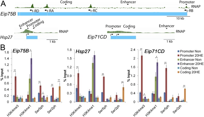
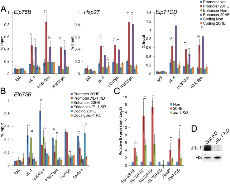
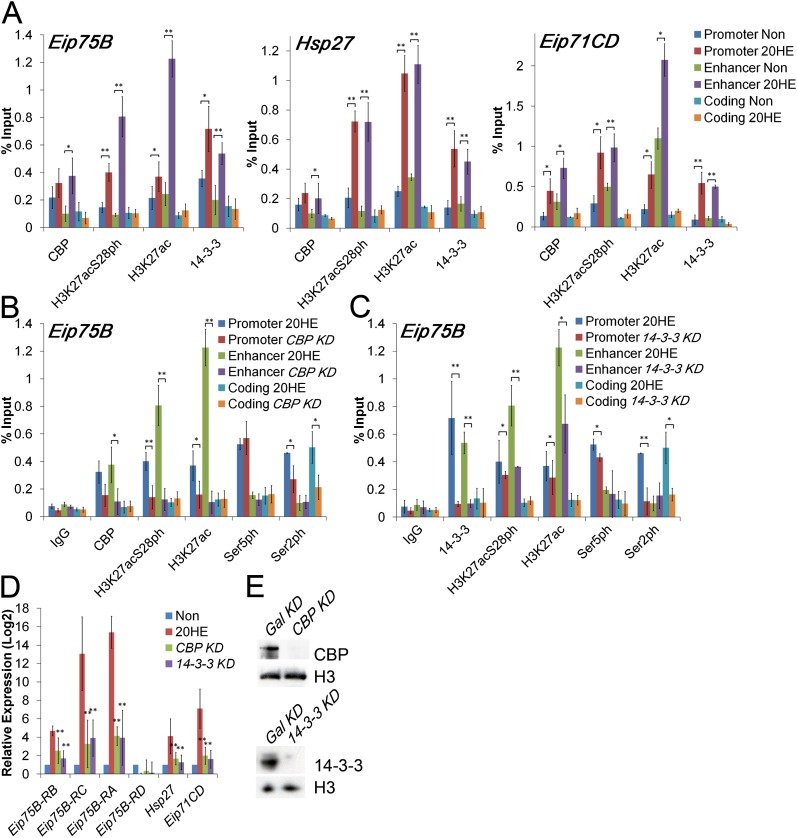
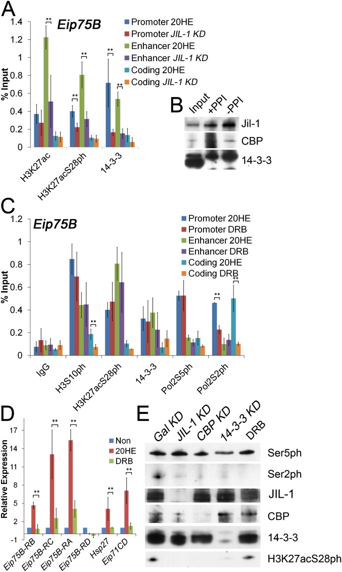
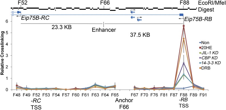
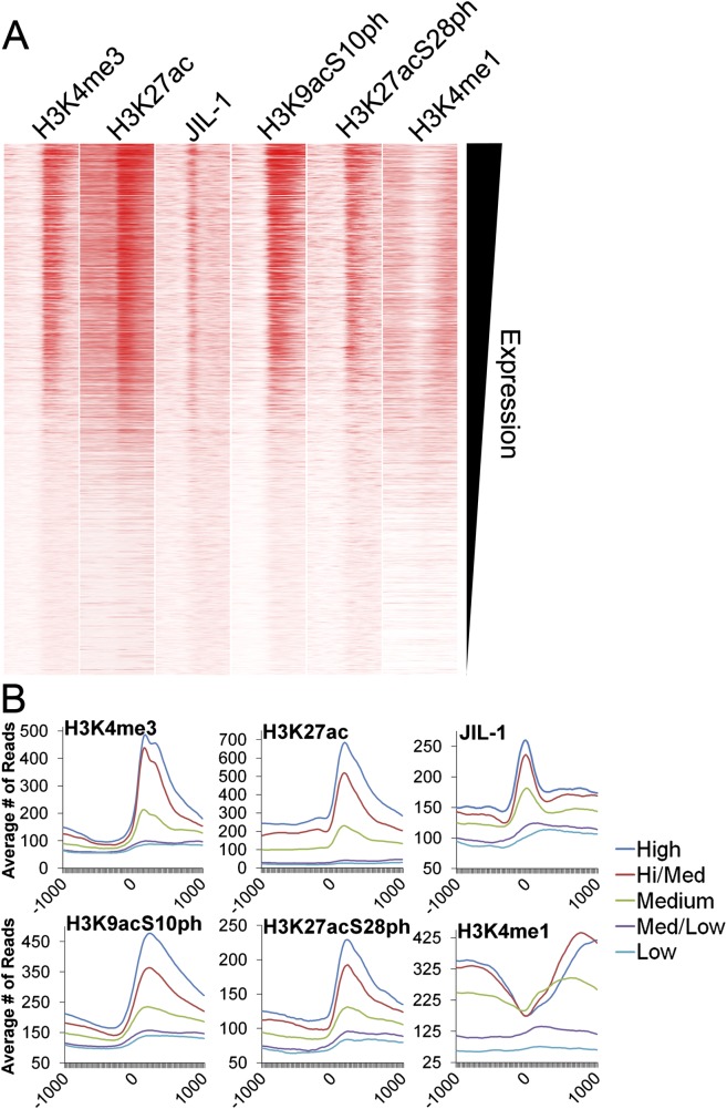
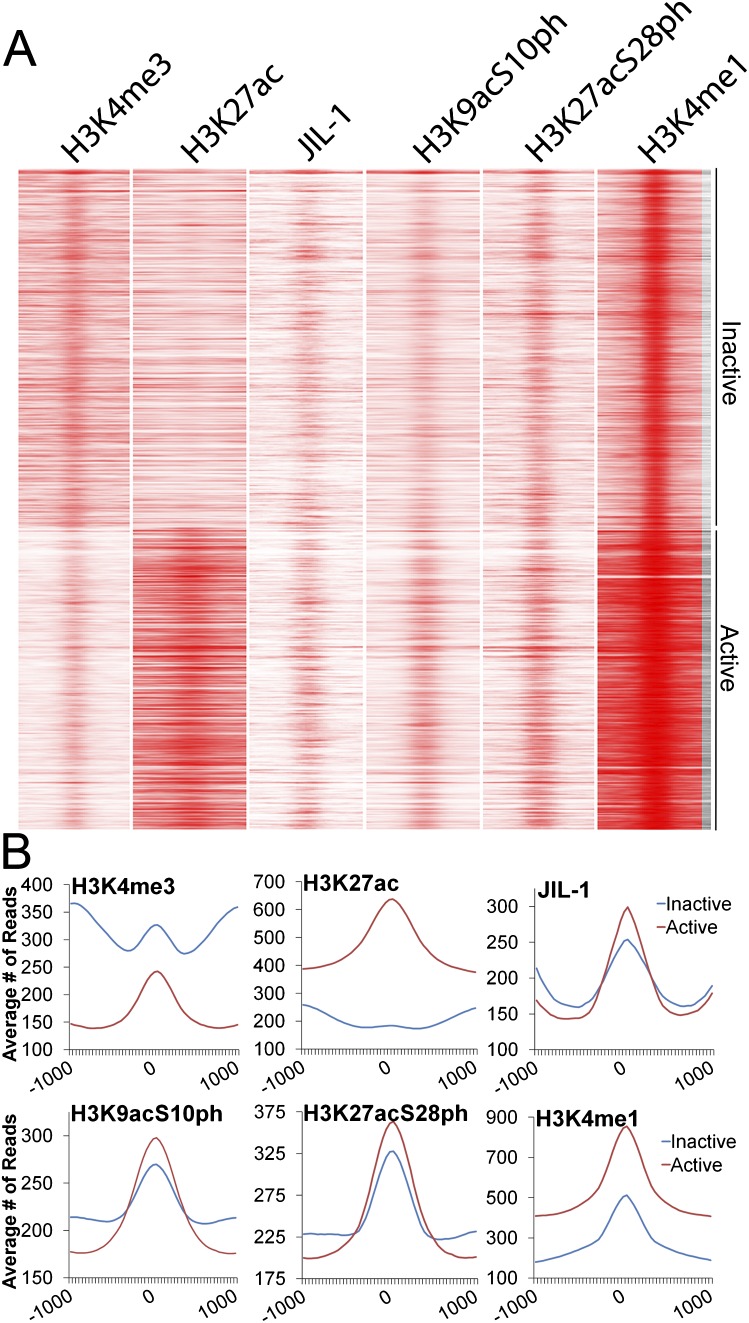
Transcription regulation is mediated by enhancers that bind sequence-specific transcription factors, which in turn interact with the promoters of the genes they control. Here, we show that the JIL-1 kinase is present at both enhancers and promoters of ecdysone-induced Drosophila genes, where it phosphorylates the Ser10 and Ser28 residues of histone H3. JIL-1 is also required for CREB binding protein (CBP)-mediated acetylation of Lys27, a well-characterized mark of active enhancers. The presence of these proteins at enhancers and promoters of ecdysone-induced genes results in the establishment of the H3K9acS10ph and H3K27acS28ph marks at both regulatory sequences. These modifications are necessary for the recruitment of 14-3-3, a scaffolding protein capable of facilitating interactions between two simultaneously bound proteins. Chromatin conformation capture assays indicate that interaction between the enhancer and the promoter is dependent on the presence of JIL-1, 14-3-3, and CBP. Genome-wide analyses extend these conclusions to most Drosophila genes, showing that the presence of JIL-1, H3K9acS10ph, and H3K27acS28ph is a general feature of enhancers and promoters in this organism.


Figures

Figure 1.
Figure 1.
Phosphorylation of H3S10 and H3S28 by JIL-1 at promoters and enhancers is necessary for transcriptional activation of ecdysone responsive genes. (A) Structure of three early 20-HE responsive genes with previously characterized enhancers showing the location of promoter, enhancer, and coding sequences used in ChIP analyses. ChIP-chip (modENCODE) profile of RNAPII large subunit indicated in green above each gene. (B) ChIP analysis of Kc167 cells treated for 3 h with ethanol (Non) or 20-HE (20HE). Antibodies used for pull-down are indicated along the x-axis. (Ser5ph) RNAPII phosphorylated on serine 5 of CTD, (Ser2ph) RNAPII phosphorylated on serine 2 of CTD. DNA was quantified by real-time PCR using primers designed to amplify the promoter, enhancer, and coding regions of each of the three genes.
Figure 2.
Figure 2.
JIL-1 is required for phosphorylation at enhancers and promoters upon ecdysone transcriptional activation. (A) ChIP analysis of Kc167 cells treated for 3 h with ethanol (Non) or 20-HE (20HE). Antibodies used for pull-down are indicated along the x-axis. (Ser5ph) RNAPII phosphorylated on serine 5 of CTD, (Ser2ph) RNAPII phosphorylated on serine 2 of CTD. DNA was quantified by real-time PCR using primers designed to amplify the promoter, enhancer, and coding regions of each of the three genes. (B) ChIP analysis in Kc167 cells in which JIL-1 or Gal expression was inhibited by RNAi. Cells were treated with 20-HE for 3 h. Antibodies used are indicated along the x-axis. (Ser5ph) RNAPII phosphorylated on serine 5 of CTD, (Ser2ph) RNAPII phosphorylated on serine 2 of CTD. DNA was quantified by real-time PCR, and results are reported as percentage of input. (C) RNA expression analysis in cells treated with dsRNAs corresponding to the JIL-1 or Gal genes. Cells were treated with ethanol (Non) or 20-HE (20HE) for 3 h. RNA levels were determined by qPCR using primers specific to each of the four Eip75B transcripts. All samples were normalized to the mitochondrial gene myt:Col and the Non sample was set to 1 for comparison. (D) Western analysis of dsRNA-treated cells showing undetectable levels of JIL-1. Error bars represent the standard deviation of the mean of 3 biological replicates. (*) P < 0.05, (**) P < 0.01.
Figure 3.
Figure 3.
CBP acetylation and 14-3-3 recruitment at promoters and enhancers is necessary for transcription activation of ecdysone-responsive genes. (A) ChIP analysis of Kc167 cells treated for 3 h with ethanol (Non) or 20-HE (20HE) was performed using the antibodies indicated along the x-axis. (Ser5ph) RNAPII phosphorylated on serine 5 of CTD, (Ser2ph) RNAPII phosphorylated on serine 2 of CTD. DNA was quantified by real-time PCR using primers designed to amplify the promoter, enhancer, and coding regions of each of the three ecdysone-induced genes. (B) ChIP analysis of Kc167 cells nontreated and treated with CBP dsRNA compared to Gal dsRNA. The cells were also incubated with 20-HE for 3 h, and chromatin was immunoprecipitated using the antibodies indicated along the x-axis. DNA was quantified by real-time PCR, and the result is reported as a percent of the input at the Eip75B gene. (C) Similar to panel B, but cells were treated with dsRNA to 14-3-3. (D) RNA expression analysis in cells treated with dsRNAs corresponding to the CBP, 14-3-3, or Gal genes. Cells were treated with ethanol (Non) or 20-HE (20HE) for 3 h. RNA levels were determined by qPCR using primers specific to each of the four Eip75B transcripts. All samples were normalized to the mitochondrial gene myt:Col and the Non sample was set to 1 for comparison. (E) Western analysis of lysates from cells treated with CBP (top) or 14-3-3 (bottom) dsRNAs. Levels of either protein are undetectable. Error bars represent the standard deviation of the mean of 3 biological replicates. (*) P < 0.05, (**) P < 0.01.
Figure 4.
Figure 4.
14-3-3 recruitment and H3K27 acetylation are dependent on JIL-1 kinase at enhancers and promoters. (A) Kc167 cells treated with dsRNAs to Gal or JIL-1 were incubated with 20-HE for 3 h and subjected to ChIP analysis using the antibodies indicated along the x-axis. DNA was quantified by real-time PCR and reported as a percent of the input. (B) Kc167 cell lysates with and without phosphatase inhibitors (PPI) were immunoprecipitated using antibodies against JIL-1, loaded 1:10 with respect to input, and subjected to Western analysis using antibodies against JIL-1, CBP, and 14-3-3. (C) Using Kc167 cells pretreated for 5 min with 100 μM DRB followed by 3-h treatment with 20-HE (20HE), ChIP was performed using the antibodies indicated along the x-axis and quantitated by real-time PCR using primers designed to amplify the promoter, enhancer, and coding regions of the Eip75B gene. (D) Relative expression analysis of the DRB-treated cells compared with normal 20-HE induction using primers specific to each of the four transcripts of Eip75B. All samples were normalized to mitochondrial gene product myt:Col and the Non sample set to 1 for comparison. Error bars represent the standard deviation of the mean of 3 biological replicates. (E) Western analysis of lysates from Kc167 cells treated with dsRNAs corresponding to the JIL-1, CBP, 14-3-3, or Gal genes; cells were pretreated with DRB for 5 min and with 20-HE for 3 h. Error bars represent the standard deviation of the mean of 3 biological replicates. (*) P < 0.05, (**) P < 0.01.
Figure 5.
Figure 5.
JIL-1, 14-3-3, and CBP are required for enhancer-promoter interactions. 3C analysis of Kc167 cells after Gal knockdown and treatment with ethanol (Non) or 20-HE (20HE) for 3 h was performed in normal cells as well as cells in which the expression of the JIL-1, CBP, 14-3-3, or Gal genes was inhibited by treatment with the corresponding dsRNAs. 3C analysis was also done on cells treated with DRB for 5 min. Cross-linking efficiencies are reported after normalizing to a BAC clone with an insert of 170 kb containing the entire Eip75B locus. A restriction digest of this DNA gives 112 fragments numbered 1 to 112, beginning with the first fragment of the BAC. Error bars represent the standard deviation of the mean of 3 biological replicates.
Figure 6.
Figure 6.
JIL-1 and histone phosphoacetylation at promoters correlate with transcript levels genome-wide. (A) RNA expression levels for Kc167 cells were obtained from modENCODE, and the TSSs were sorted by expression level from highest to lowest and given up/downstream orientation according to strand. The total number of reads is plotted for each ChIP-seq data set in 20-bp bins covering 1 kb upstream of and downstream from each TSS and viewed in Java Treeview. (B) All TSSs were broken down into five equal bins according to expression levels, and the average number of reads for each 20-bp region for all the TSSs in that bin is plotted 1 kb upstream and downstream for each bin.
Figure 7.
Figure 7.
JIL-1, H3K9acS10ph, and H3K27acS28ph are present at enhancers. Enhancers were selected by the presence of H3K4m1 and clustered into two clusters using Cluster 3.0. (A) The total number of reads is plotted for each ChIP-seq data set in 20-bp bins covering 1 kb upstream and downstream from the H3K4me1 peak summit and viewed in Java Treeview. (B) The average number of reads plotted for every H3K4me1 peak in the inactive or active cluster in 20-bp bins covering 1 kb upstream and downstream from the summit of H3K4me1.

Similar articles

Cited by

References

    1. Berger SL 2007. The complex language of chromatin regulation during transcription. Nature 447: 407–412 - PubMed
    1. Bernardo TJ, Dubrovskaya VA, Jannat H, Maughan B, Dubrovsky EB 2009. Hormonal regulation of the E75 gene in Drosophila: Identifying functional regulatory elements through computational and biological analysis. J Mol Biol 387: 794–808 - PubMed
    1. Cai W, Bao X, Deng H, Jin Y, Girton J, Johansen J, Johansen KM 2008. Pol II-mediated transcription at active loci does not require histone H3S10 phosphorylation in Drosophila. Development 135: 2917–2925 - PMC - PubMed
    1. Cherbas L, Lee K, Cherbas P 1991. Identification of ecdysone response elements by analysis of the Drosophila Eip28/29 gene. Genes Dev 5: 120–131 - PubMed
    1. Choudhary C, Kumar C, Gnad F, Nielsen ML, Rehman M, Walther TC, Olsen JV, Mann M 2009. Lysine acetylation targets protein complexes and co-regulates major cellular functions. Science 325: 834–840 - PubMed

Publication types

MeSH terms

Associated data