Case Reports
. 2012 Jul;80(7):2346-53.
doi: 10.1128/IAI.00293-12. Epub 2012 Apr 16.

Invasive potential of nonencapsulated disease isolates of Neisseria meningitidis

Affiliations
Case Reports

Invasive potential of nonencapsulated disease isolates of Neisseria meningitidis

Kay O Johswich et al. Infect Immun. 2012 Jul.

Abstract

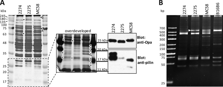
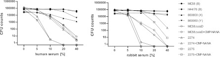
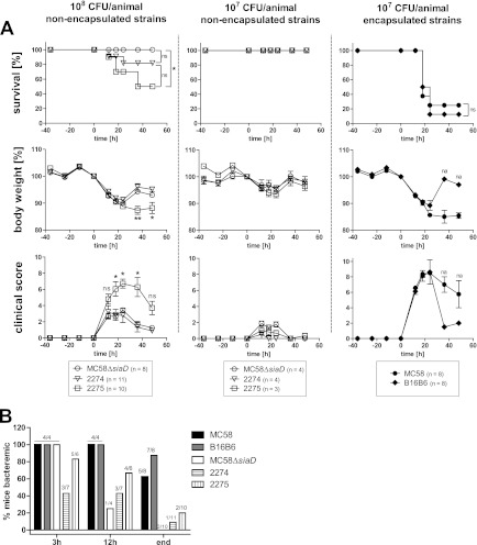
The capsule of Neisseria meningitidis is the major virulence factor that enables this bacterium to overcome host immunity elicited by complement and phagocytes, rendering it capable of surviving in blood. As such, nonencapsulated N. meningitidis isolates are generally considered nonpathogenic. Here, we consider the inherent virulence of two nonencapsulated N. meningitidis isolates obtained from our national surveillance of infected blood cultures in Canada. Capsule deficiency of both strains was confirmed by serology and PCR for the ctrA to ctrD genes and siaA to siaC genes, as well as siaD genes specific to serogroups B, C, Y, and W135. In both strains, the capsule synthesis genes were replaced by the capsule null locus, cnl-2. In accordance with a lack of capsule, both strains were fully susceptible to killing by both human and baby rabbit complement. However, in the presence of cytidine-5' monophospho-N-acetylneuraminic acid (CMP-NANA), allowing for lipooligosaccharide (LOS) sialylation, a significant increase of resistance to complement killing was observed. Mass spectrometry of purified LOS did not reveal any uncommon modifications that would explain their invasive phenotype. Finally, in a mouse intraperitoneal challenge model, these nonencapsulated isolates displayed enhanced virulence relative to an isogenic mutant of serogroup B strain MC58 lacking capsule (MC58ΔsiaD). Virulence of all nonencapsulated isolates tested was below that of encapsulated serogroup B strains MC58 and B16B6. However, whereas no mortality was observed with MC58ΔsiaD, 5/10 mice succumbed to infection with strain 2275 and 2/11 mice succumbed to strain 2274. Our results suggest the acquisition of a new virulence phenotype by these nonencapsulated strains.


Figures

Fig 1
Fig 1
Phenotypic characterization of nonencapsulated N. meningitidis strains. (A) Whole-cell lysates of strains 2274, 2275, and MC58 were analyzed on a 12% polyacrylamide gel and visualized by silver staining (left). The indicated low-molecular-mass portion of the gel after prolonged silver stain development is shown to the right. Differences in band intensities or band presence are indicated by arrowheads. Black arrowheads indicate the presence of a protein band missing or expressed at a significantly different level or with a different molecular mass than in the other strain(s). White arrowheads indicate protein bands missing or significantly fainter in MC58 than in 2274 and 2275. Immunoblots for Opa and pilin are shown on the right (note that the molecular mass markers are aligned with those of the adjacent [overdeveloped] silver-stained gel.) (B) PCR-based opa typing of strains 2274, 2275, MC58, and B16B6. The white arrowhead indicates a band that was detected in strain 2275 but not 2274.
Fig 2
Fig 2
Relative susceptibility of N. meningitidis isolates to killing with human versus rabbit serum as the source of complement. A total of ∼105 CFU of each strain was exposed to dilutions of serum for 30 min at 37°C in minimal essential medium, and then serial dilutions were plated onto GC agar plates to assess viable CFU counts. Nonencapsulated strains 2274 and 2275 as well as the capsule-deficient strain MC58ΔsiaD, grown in the presence or absence of 20 μM CMP-NANA, were compared to encapsulated clinical isolates MC58 (serogroup B), H44/76 (serogroup B), 86800 (serogroup X), and 860060 (serogroup Y).
Fig 3
Fig 3
The mouse intraperitoneal infection model of invasive meningococcal disease. (A) Mice were infected with 107 CFU of encapsulated N. meningitidis strain MC58 or B16B6 (right panels) or with 108 or 107 CFU of N. meningitidis MC58ΔsiaD (capsule-deficient mutant) or the nonencapsulated clinical isolates strain 2274 and strain 2275 (center and left). From 36 h prior to infection until 48 h after infection, mice were monitored every 12 h (and also at 18 h postinfection) for survival (upper panels), loss of body weight (middle panel), and clinical scores, assigned to reflect morbidity (lower panels). Statistic analyses were performed using GraphPad Prism 5. The Mantel-Cox log rank test was used for survival analysis (*, P < 0.05). A one-way analysis of variance applying Bonferroni's post hoc test was used for statistical analysis of body weight (*, P < 0.05, **, P < 0.01). The Kruskal-Wallis test with application of Dunn's post hoc test was used to analyze clinical scoring data (*, P < 0.05). For body weight and clinical score data, only the lowest significance levels of differences between strain 2275 and the other two groups are indicated. na, no statistics applicable because only one (B16B6) or two (MC58) animals survived. (B) Assessment of bacteremia in intraperitoneally infected mice. Tail vein blood samples were drawn at the indicated times after infection of mice and plated to assess viable bacteria in the peripheral blood. Depicted is the percentage of mice testing culture positive for N. meningitidis. Numbers on top of columns indicate the numbers of culture-positive animals among the total number of tested animals.

References

    1. Abdillahi H, Poolman JT. 1987. Whole-cell ELISA for typing Neisseria meningitidis with monoclonal antibodies. FEMS Microbiol. Lett. 48:367–371 - PubMed
    1. Ashton FE, Ryan A, Diena BB, Frasch CE. 1979. Evaluation of the antiserum agar method for the serogroup identification of Neisseria meningitidis. Can. J. Microbiol. 25:784–787 - PubMed
    1. Bennett DE, Mulhall RM, Cafferkey MT. 2004. PCR-based assay for detection of Neisseria meningitidis capsular serogroups 29E, X, and Z. J. Clin. Microbiol. 42:1764–1765 - PMC - PubMed
    1. Borrow R, et al. 1998. siaD PCR ELISA for confirmation and identification of serogroup Y and W135 meningococcal infections. FEMS Microbiol. Lett. 159:209–214 - PubMed
    1. Borrow R, et al. 1997. Non-culture diagnosis and serogroup determination of meningococcal B and C infection by a sialyltransferase (siaD) PCR ELISA. Epidemiol. Infect. 118:111–117 - PMC - PubMed

Publication types

MeSH terms

Substances