. 2012 Jul;32(13):2467-78.
doi: 10.1128/MCB.06633-11. Epub 2012 Apr 16.

The extracellular signal-regulated kinase 3 (mitogen-activated protein kinase 6 [MAPK6])-MAPK-activated protein kinase 5 signaling complex regulates septin function and dendrite morphology

Affiliations

The extracellular signal-regulated kinase 3 (mitogen-activated protein kinase 6 [MAPK6])-MAPK-activated protein kinase 5 signaling complex regulates septin function and dendrite morphology

Frank Brand et al. Mol Cell Biol. 2012 Jul.

Abstract

Mitogen-activated protein kinase-activated protein (MAPKAP) kinase 5 (MK5) deficiency is associated with reduced extracellular signal-regulated kinase 3 (ERK3) (mitogen-activated protein kinase 6) levels, hence we utilized the MK5 knockout mouse model to analyze the physiological functions of the ERK3/MK5 signaling module. MK5-deficient mice displayed impaired dendritic spine formation in mouse hippocampal neurons in vivo. We performed large-scale interaction screens to understand the neuronal functions of the ERK3/MK5 pathway and identified septin7 (Sept7) as a novel interacting partner of ERK3. ERK3/MK5/Sept7 form a ternary complex, which can phosphorylate the Sept7 regulators Binders of Rho GTPases (Borgs). In addition, the brain-specific nucleotide exchange factor kalirin-7 (Kal7) was identified as an MK5 interaction partner and substrate protein. In transfected primary neurons, Sept7-dependent dendrite development and spine formation are stimulated by the ERK3/MK5 module. Thus, the regulation of neuronal morphogenesis is proposed as the first physiological function of the ERK3/MK5 signaling module.


Figures

Fig 1
Fig 1
Determination of spine numbers for MK5 deficiency in mice in vivo. (A) To visualize hippocampal neurons, Golgi staining was performed with 150-μm vibratome sections of three age-matched WT and MK5−/− mice each. Representative images for each of the animals are shown. (B) Statistical analysis of spine number from three animals of each genotype by one-tailed t test. The numbers of dendrites analyzed were 78 (WT) and 95 (MK5 knockout). (C) Analysis of dendritic spines from WT and MK5−/− animals expressing neuron-specific Thy1-YFP reporter protein. Representative images for each of the animals are shown. (D) Statistical analysis of spine number from three animals of each genotype. The numbers of dendrites analyzed were 131 (WT) and 162 (MK5 knockout).
Fig 2
Fig 2
Characterization of the ERK3-Sept7 interaction. (A) Schematic representation of deletion constructs of Sept7 generated for pulldown studies (PR, proline rich; CC, coiled-coil domain). (B) Scheme representing the ERK3 deletion mutants. The activating phosphorylation site is indicated. (C) GST-tagged deletion mutants of Sept7, which lack either or both the C and N termini, were used in a GST pulldown (PD) of BE-tagged ERK3. Only Sept7 with the complete C terminus (CT) can bind to ERK3. IB, immunoblot. SA, streptavidin detection of Bio-Ease tag. (D) Pulldown of BE-tagged Sept7ΔN with GST-tagged C-terminal deletion mutants of ERK3. The region of ERK3 between amino acids 358 and 720 (ΔC2) is essential for effective interaction with Sept7ΔN. (E) Pulldown of endogenous ERK3 by overexpressed GST-Sept7, but not by GST-Sept7ΔC, from lysates of HEK293 cells. (F) Pulldown of endogenous Sept7 by GST-ERK3 from lysates of HEK293 cells. The level of Sept7 in the pulldown is reduced when GST-ERK3ΔC3 is used.
Fig 3
Fig 3
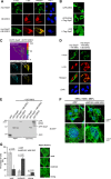
ERK3 acts as a docking platform for Sept7 and MK5. (A) Coexpression of BE-tagged ERK3 with myc-tagged Sept7 leads to the subcellular relocalization of ERK3 to Sept7 filament in HEK293 cells. (B) Subcellular localization of GFP-ERK3 is altered by the coexpression of Flag-tagged Sept7 in HeLa cells from uniform cytoplasmic localization to filamentous cytoplasmic structures. GFP transfected with Flag-Sept7 is shown as a control (scale bar, 5 μm). (C) FRET measurements of the interaction of ECFP-Sept7 and EYFP-ERK3 in living HeLa cells by acceptor photobleaching. FRET efficiency (FRET Eff) is calculated from the donor fluorescence before (D pre) and after bleaching of the acceptor (D post) using the equation FRET Eff = 1 (D pre/D post), and results are shown by false color coding. The fluorescence of the donor and the acceptor (A) before and after bleaching are shown separately. (D) MK5 can bind to Sept7-bound ERK3 and relocalize to Sept7 filamentous structures (scale bar, 5 μm). (E) MK5 interaction with Sept7 is bridged by ERK3 binding. GST pulldown assay with GST-MK5 in HEK293 cells showed copurification of GFP-Sept7 only if ERK3 was also coexpressed. (F) Endogenous Sept7 filaments are less organized in ERK3/MK5 double-knockout MEFs. ERK3/MK5 knockout MEFs retrovirally rescued with ERK3 and MK5 (pMMP-MK5/pMMP-ERK3) showed a higher complexity of Sept7-containing filaments than the empty vector-transduced cells (pMMP). (G) Categorization of septin filament complexity performed for 100 of the differentially transduced cells in triplicate (standard deviations are indicated) into groups of weak, moderate, or strong filament organization. ERK3/MK5 retrovirally rescued cells showed a significantly higher number of moderate and strong septin filament-containing cells and a significantly lower number of cells displaying weak septin organization (one-tailed t test).
Fig 4
Fig 4
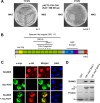
Identification and characterization of Kal7 as a novel MK5 interaction partner. (A) Identification of RhoGEF kalirin-7 (Kal7) as an MK5 interacting protein in a classical Y2H screen. pGBKT7-MK5 was used as bait for the screening of a brain cDNA library (Clontech). A cDNA clone expressing a 353-amino-acid fragment of Kal7 specifically interacts with MK5 but not with the related protein kinase MK2 or the empty bait vector. (B) Schematic representation of the domain structure and the MK5 interacting region of mouse Kal7 (DH, Dbl homology; GEF, guanosine nucleotide exchange factor; PH, pleckstrin homology; PDZ, PDZ binding motif [Ser-Thr-Tyr-Val]; sec14p, domain binding PIP2 [3, 5] and PIP3; and SR, spectrin-like repeat). (C) myc-Kal7 and HA-MK5 were coexpressed in HEK293 cells and analyzed by immunofluorescence. DAPI staining shows the nuclei in blue. (D) Pulldown of endogenous MK5 from HEK293 cells using GST-tagged fragments of Kal7 spanning spectrin-like repeats 3 to 6 or 4 to 7.
Fig 5
Fig 5
Borg1 to Borg3 and Kal7 as in vitro substrates of ERK3 and MK5. (A) In vitro phosphorylation of Borg1, Borg2, and Borg3 by ERK3 and MK5. Recombinant GST-tagged Borg1, Borg2, and Borg3 were expressed in E. coli and used for in vitro kinase assays with Sf9-expressed, active His-ERK3 or with E. coli-expressed, p38-activated GST-MK5. Incorporated phosphate from [γ-33P]ATP was detected by phosphorimaging. As positive controls, the known ERK3 substrate myelin basic protein (MBP) and the known MK5 substrate heat shock protein beta-1 (Hspb1; Hsp25) were used. ERK3 phosphorylates all Borg proteins, especially Borg1 and Borg2, whereas MK5 preferentially phosphorylates Borg2. This correlates with the existence of phosphorylation site consensus motifs identified in these proteins. (B) Alignment of MK5 consensus phosphorylation motif in Kal7 to that in the in vitro substrate Hspb1. Kal7-S 487 is located in an ideal MK5 consensus motif, LXRXXS*, and a putative SH3-binding PXS*P motif. (C) Identification of the MK5 phosphorylation site in Kal7. The fragment SR3-6, but not the mutated fragment SR3-6-S487A, is phosphorylated by MK5. Phosphorylation was detected by 32P incorporation and phosphorimaging.
Fig 6
Fig 6
Sept7-dependent stimulation of dendritic outgrowth and increased spine number by overexpression of the ERK3/MK5 complex in primary hippocampal neurons. Primary neurons were isolated at embryonic day 18. Transfection was carried out with BE-tagged ERK3, HA-tagged MK5, myc-tagged Sept7, or their combinations together with pEGFP at DIV5 (A to C) or DIV10 (D). Seventy-two (A to C) or 120 (D) h posttransfection, cells were fixed and analyzed using confocal microscopy. (A) Representative images of statistically relevant neurons overexpressing GFP (CTRL), Sept7, or a combination of ERK3, MK5, and Sept7 (scale bar, 50 μm). (B) Sholl analysis of neurons differently transfected with the constructs coding for the indicated proteins and EGFP. (C) Qualitative analysis of dendritic spine morphology from the experiment shown in panel B. (D) Representative images of primary neurons expressing EGFP and Sept7 or ERK3/MK5/Sept7, respectively (scale bar, 50 μm). Insets display spine morphology.
Fig 7
Fig 7
Neuronal ERK3/MK5/Sept7 signaling and spine morphogenesis. The ERK3/MK5/Sept7 signaling complex is activated by Cdc42 and Rac GTPase via p21-activated protein kinases (PAKs). Septin heteromeric structures are generated via the ERK3/MK5/Sept7-mediated recruitment and phosphorylation of Borg proteins. The MK5 substrate Kal7, a Rho GEF and known activator of Rac GTPases, further contributes to PAK activation and actin filament reorganization. Thus, the coordinated phosphorylation of Borg proteins and Kal7 by ERK3 and MK5 constitute a novel signaling cascade involving feed-forward circuits, multiple GTPases, and cytoskeletal elements.

Similar articles

Cited by

References

    1. Aberg E, et al. 2006. Regulation of MAPK-activated protein kinase 5 activity and subcellular localization by the atypical MAPK ERK4/MAPK4. J. Biol. Chem. 281: 35499–35510 - PubMed
    1. Aberg E, et al. 2009. Docking of PRAK/MK5 to the atypical MAPKs ERK3 and ERK4 defines a novel MAPK interaction motif. J. Biol. Chem. 284: 19392–19401 - PMC - PubMed
    1. Barral Y. 2010. Cell biology. Septins at the nexus. Science 329: 1289–1290 - PubMed
    1. Barral Y, Mansuy IM. 2007. Septins: cellular and functional barriers of neuronal activity. Curr. Biol. 17: R961–R963 - PubMed
    1. Broder YC, Katz S, Aronheim A. 1998. The ras recruitment system, a novel approach to the study of protein-protein interactions. Curr. Biol. 8: 1121–1124 - PubMed

Publication types

MeSH terms