. 2012 May 1;109(18):7037-42.
doi: 10.1073/pnas.1202288109. Epub 2012 Apr 16.

Long-lived epithelial immunity by tissue-resident memory T (TRM) cells in the absence of persisting local antigen presentation

Affiliations

Long-lived epithelial immunity by tissue-resident memory T (TRM) cells in the absence of persisting local antigen presentation

Laura K Mackay et al. Proc Natl Acad Sci U S A. .

Abstract

Although circulating memory T cells provide enhanced protection against pathogen challenge, they often fail to do so if infection is localized to peripheral or extralymphoid compartments. In those cases, it is T cells already resident at the site of virus challenge that offer superior immune protection. These tissue-resident memory T (T(RM)) cells are identified by their expression of the α-chain from the integrin α(E)(CD103)β(7), and can exist in disequilibrium with the blood, remaining in the local environment long after peripheral infections subside. In this study, we demonstrate that long-lived intraepithelial CD103(+)CD8(+) T(RM) cells can be generated in the absence of in situ antigen recognition. Local inflammation in skin and mucosa alone resulted in enhanced recruitment of effector populations and their conversion to the T(RM) phenotype. The CD8(+) T(RM) cells lodged in these barrier tissues provided long-lived protection against local challenge with herpes simplex virus in skin and vagina challenge models, and were clearly superior to the circulating memory T-cell cohort. The results demonstrate that peripheral T(RM) cells can be generated and survive in the absence of local antigen presentation and provide a powerful means of achieving immune protection against peripheral infection.


Conflict of interest statement

The authors declare no conflict of interest.

Figures

Fig. 1.
Fig. 1.
Circulating memory CD8+ T cells fail to provide efficient protection against HSV-1 skin infection. (A and B) Mice were immunized by intranasal flu.gB and infected with HSV during the effector (day 10) or memory (day 30) phase of the flu.gB response. A control cohort was not immunized (i.e., naive). (A) Viral titres in the skin 3 d after HSV infection and (B) representative photographs show lesion formation 6 d following HSV infection. (C and D) Mice were seeded with naive gBT-I T cells, and T cells were primed with gB peptide-pulsed DCs, followed by flu.gB intranasal booster infection 1 wk (days 7–8, n = 7 per group), 2 wk (days 12–19, n = 9–12 per group), or 9 to 13 wk (days 63–89, n = 9–10 per group) after immunization. (C) Frequency of gBT-I memory cells in peripheral blood during the effector and memory phases of flu.gB infection (days 10 and 30, respectively) or following DC priming and subsequent flu.gB infection [DC(gB)+flu.gB] or control immunization [DC(no)+flu.gB], 1 d before HSV challenge infection. Bars represent mean ± SEM. (D) Viral titers in the skin 6 d following HSV infection. Symbols represent individual mice; bars represent the mean. Data are pooled from five independent experiments.
Fig. 2.
Fig. 2.
T-cell persistence in skin sites subject to nonspecific inflammation. Mice were seeded with in vitro activated gBT-I T cells and treated with DNFB on the left flank. (A) Number of gBT-I T cells in DNFB-treated or nontreated control (ctrl) skin sites at the indicated times after treatment. Bars represent mean ± SEM (n = 4–8 per group). (B) Representative flow cytometry plots showing gBT-I T cells in DNFB-treated or control skin sites and in the spleen 360 d after DNFB treatment (n = 5–6).
Fig. 3.
Fig. 3.
CD103 is selectively up-regulated on T cells in skin in the absence of local antigen stimulation. (A) Representative flow cytometry plot showing CD103 expression by gBT-I T cells in DNFB-treated skin and the spleen 360 d after DNFB treatment. (B) Mice were seeded with naive gBT-I T cells and 1 × 10⁷ in vitro activated OT-I T cells and infected with HSV KOS (WT virus) on the left flank and the HSV variant K.L8A on the right flank of the same mouse. Shown is CD103 expression by gBT-I T cells in the skin and ganglia of HSV KOS-infected flanks (gBT-I/KOS; black line), gBT-I T cells in the skin and ganglia of flanks infected with the HSV variant K.L8A (gBT-I/K.L8A; dotted line), and OT-I T cells in the skin and ganglia of HSV KOS-infected flanks (OT-I; gray filled line), at days 28 to 35 after infection. All data are representative of three independent experiments.
Fig. 4.
Fig. 4.
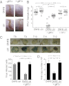
Virus-specific T cells artificially lodged in the skin are protective against infection with HSV. Mice were seeded with in vitro-activated gBT-I T cells and treated with DNFB on the left flank. A control cohort did not receive gBT-I T cells or received in vitro-activated OT-I T cells. Mice were infected with HSV on both left (DNFB) and right (ctrl) skin flanks 30 to 38 d after DNFB treatment. (A) Representative photographs showing lesion formation and (B) viral titers in the skin 6 d after HSV infection in the cohorts described. Symbols represent individual mice; bars represent the mean. Data are pooled from three experiments. (C) Mice were treated as described and infected on the left and right flanks with HSV KOS.LβA virus at day 35 after DNFB treatment. At day 20 after infection, the innervating ganglia (T8–12) were harvested, fixed, and stained to detect β-gal expression in infected cells. Shown are representative photomicrographs of whole mounted ganglia from a single mouse (Left), and pooled data are shown in the bar graph. Bars represent mean ± SEM of viral-infected cells in pooled ganglia/mouse (n = 5 mice per group). (D) Viral copy number in the innervating ganglia at day 20 after HSV infection. Bars represent mean ± SEM. Data are representative of two independent experiments.
Fig. 5.
Fig. 5.
TRM cells offer superior protection against HSV compared with circulating memory T cells. Mice were seeded with in vitro-activated gBT-I T cells at the time of DNFB treatment or 15 d before treatment. Shown is the number of gBT-I T cells in (A) DNFB-treated skin sites and (B) the spleen 10 d after treatment. Bars represent mean ± SEM. (C) Cohorts of mice were infected with HSV on both left and right flanks 30 d following DNFB treatment. Shown are viral titers in the skin 6 d after HSV infection. Symbols represent individual mice; bars represent the mean. Data are representative of two experiments.
Fig. 6.
Fig. 6.
TRM cells can protect against HSV challenge in different tissues and after different lodgement modalities. (A and B) Mice were seeded with naive gBT-I T cells and immunized with flu.gB intranasally. A control cohort was nonimmunized. Mice were treated with DNFB on the left flank 10 d after infection. (A) Number of gBT-I T cells in DNFB-treated skin in cohorts with and without flu.gB infection 30 d after DNFB treatment. Bars represent mean ± SEM. (B) Mice were infected with HSV on left (DNFB) and right (ctrl) skin flanks 30 d after DNFB treatment. Shown are viral titers in the skin at day 6 after infection. Symbols represent individual mice; bars represent the mean. Data are pooled from two experiments. (C and D) Mice were treated intravaginally with N9 and transferred with in vitro-activated gBT-I T cells at day 2 of treatment. (C) Number of gBT-I T cells in the vagina of N9-treated or nontreated mice 30 d following treatment. Bars represent mean ± SEM (n = 3–4 per group) and are representative of three experiments. (D) Mice were infected intravaginally with HSV 30 d after N9 treatment (N9 + gBT-I). Control cohorts were N9-treated mice without gBT-I T cells transferred (N9) and mice with gBT-I T-cell transferred that were not N9-treated (gBT-I). Shown are viral titres from vaginal swabs at the indicated days after infection. Symbols represent individual mice; bars represent the mean. Data are pooled from three experiments.

References

    1. Welsh RM, Selin LK, Szomolanyi-Tsuda E. Immunological memory to viral infections. Annu Rev Immunol. 2004;22:711–743. - PubMed
    1. Harty JT, Tvinnereim AR, White DW. CD8+ T cell effector mechanisms in resistance to infection. Annu Rev Immunol. 2000;18:275–308. - PubMed
    1. Bachmann MF, Kündig TM, Hengartner H, Zinkernagel RM. Protection against immunopathological consequences of a viral infection by activated but not resting cytotoxic T cells: T cell memory without “memory T cells”? Proc Natl Acad Sci USA. 1997;94:640–645. - PMC - PubMed
    1. Bachmann MF, Wolint P, Schwarz K, Oxenius A. Recall proliferation potential of memory CD8+ T cells and antiviral protection. J Immunol. 2005;175:4677–4685. - PubMed
    1. Badovinac VP, Messingham KA, Jabbari A, Haring JS, Harty JT. Accelerated CD8+ T-cell memory and prime-boost response after dendritic-cell vaccination. Nat Med. 2005;11:748–756. - PubMed

Publication types

MeSH terms