. 2012 Apr 7;18(13):1485-95.
doi: 10.3748/wjg.v18.i13.1485.

HBx activates FasL and mediates HepG2 cell apoptosis through MLK3-MKK7-JNKs signal module

Affiliations

HBx activates FasL and mediates HepG2 cell apoptosis through MLK3-MKK7-JNKs signal module

Ren-Xian Tang et al. World J Gastroenterol. .

Abstract

Aim: To investigate the possible mechanism by which hepatitis B virus X protein (HBx) mediates apoptosis of HepG2 cells.

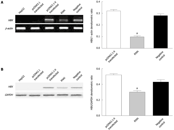
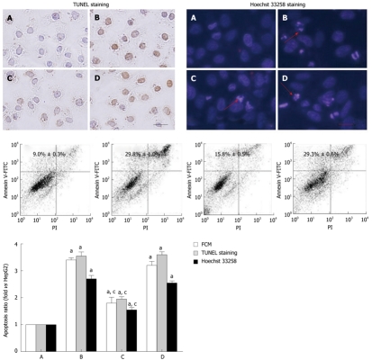
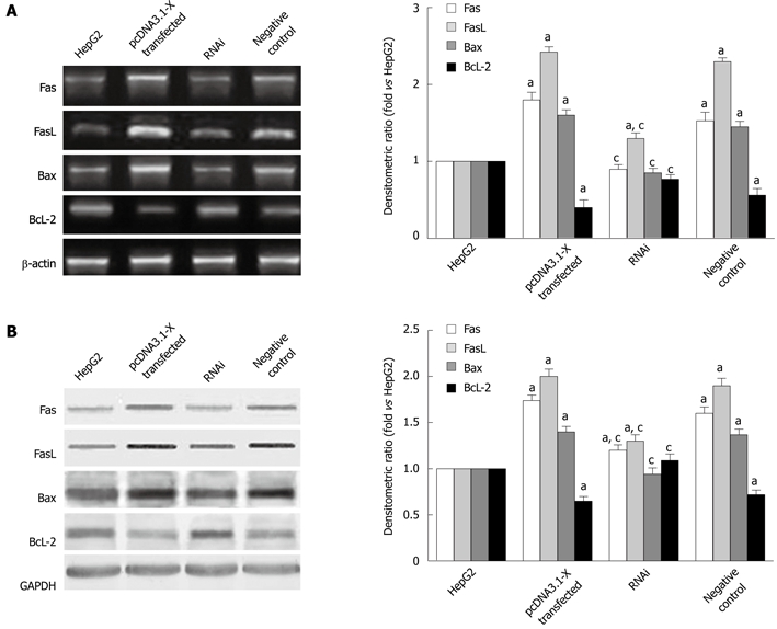
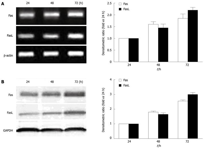
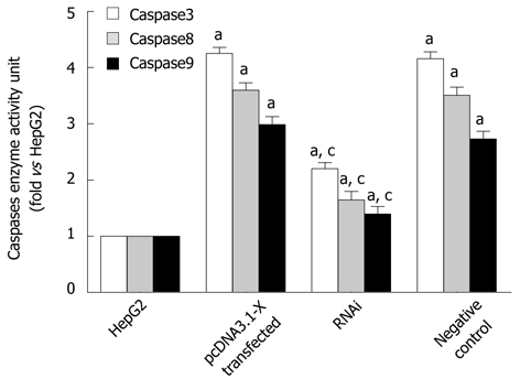
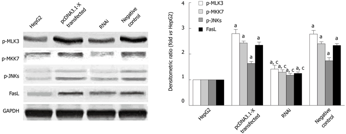
Methods: HBx expression vector pcDNA3.1-X was transfected into HepG2 cells to establish an HBx high-expression cellular model as pcDNA3.1-X transfected group. The pcDNA3.1-X and pSilencer3.1-shHBX (HBx antagonist) were cotransfected into HepG2 cells to establish an HBx low-expression model as RNAi group. Untransfected HepG2 cells and HepG2 cells transfected with negative control plasmid were used as controls. Apoptosis rate, the expression of Fas/FasL signaling pathway-related proteins and the phosphorylation levels of MLK3, MKK7 and JNKs, which are upstream molecules of death receptor pathways and belong to the family of mitogen-activated protein kinases (MAPKs), were measured in each group.

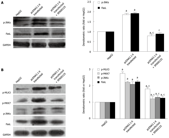
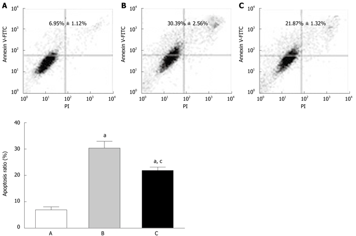
Results: Compared with HepG2 cell group and RNAi group, apoptosis rate, the expression of Fas and FasL proteins, and the activation of MLK3, MKK7 and JNKs were increased in the pcDNA3.1-X transfected group. The activation of JNKs and expression of FasL protein were inhibited in the pcDNA3.1-X transfected group when treated with a known JNK inhibitor, SP600125. When authors treated pcDNA3.1-X transfected group with K252a, a known MLK3 inhibitor, the activation of MLK3, MKK7 and JNKs as well as expression of FasL protein was inhibited. Furthermore, cell apoptosis rate was also significantly declined in the presence of K252a in the pcDNA3.1-X transfected group.

Conclusion: HBx can induce HepG2 cell apoptosis via a novel active MLK3-MKK7-JNKs signaling module to upregulate FasL protein expression.

Keywords: Apoptosis; FasL; HepG2 cell; Hepatitis B virus X protein; MLK3.


Figures

Figure 1
Figure 1
Detection of hepatitis B virus X protein expression in transfected HepG2 cells. HepG2 cells were transfected with pcDNA3.1-X plasmids or cotransfected with pcDNA3.1-X and pSilencer3.1-shHBX plasmids. Forty-eight hours later, the expression of hepatitis B virus X protein (HBx) in HepG2 cells was determined by reverse transcription polymerase chain reaction (A) and Western blotting analysis (B). HepG2 group was not transfected with any plasmids. pcDNA3.1 transfected group was transfected with plasmid pcDNA3.1; pcDNA3.1-X transfected group was transfected with pcDNA3.1-X; RNAi group was cotransfected with pcDNA3.1-X and pSilencer3.1-shHBX in a ratio of 1:3; negative control group was cotransfected with pcDNA3.1-X and negative control plasmid pSilencer3.1-H1 in a ratio of 1:3. Data are expressed as mean ± SD (n = 3), aP < 0.05 vs pcDNA3.1-X transfected group.
Figure 2
Figure 2
Apoptosis of HepG2 cells induced by hepatitis B virus X protein. HepG2 cells were cotransfected with pcDNA3.1-X and pSilencer3.1-shHBX as RNAi group, and HepG2 cells were cotransfected with pcDNA3.1-X and pSilencer3.1-H1 plasmids as negative control. Cells were examined by TUNEL, Hoechst 33258 staining and flow cytometry as described in Materials and Methods. A: HepG2 group; B: pcDNA3.1-X transfected group; C: RNAi group; D: Negative control group. Data was expressed as mean ± SD (n = 3), aP < 0.05 vs the HepG2 group; cP < 0.05 vs pcDNA3.1-X transfected group and negative control group. Scale bar value: 5 μm. Red arrows indicate apoptotic nuclei. Apoptosis ratio= (apoptotic cells/total cells) × 100%. TUNEL: Terminal deoxynucleotidyl transferase-mediated dUTP-biotin nick end labeling.
Figure 3
Figure 3
Expression of Fas, FasL, Bax, Bcl-2 mRNA and protein in HepG2 cells induced by hepatitis B virus X protein. Transfection of HepG2 cells is described in Figure 2. Forty-eight hours after transfection, the mRNA (A) and protein (B) expression levels of Bax, Bcl-2, Fas and FasL were determined by RT-PCR and Western blotting analysis. Data are expressed as mean ± SD (n = 3), aP < 0.05 vs the HepG2 group; cP < 0.05 vs pcDNA3.1-X transfected group and negative control group.
Figure 4
Figure 4
Upregulation of Fas/FasL signaling pathway-related protein expression by hepatitis B virus X protein in a time-dependent manner. HepG2 cells were transfected with pcDNA3.1-X and incubated for various time periods as indicated. The levels of Fas, FasL mRNA (A) and proteins (B) were determined by reverse transcription polymerase chain reaction and Western blotting analysis. Data are expressed as mean ± SD (n = 3).
Figure 5
Figure 5
Detection of activated caspases in HepG2 cells transfected with hepatitis B virus X protein. Transfection of HepG2 cells is described in Figure 2. Forty-eight hours after transfection, the enzyme activity of caspase3, caspase8 and caspase9 was analyzed by spectrophotometric test. Data are expressed as mean ± SD (n = 3), aP < 0.05 vs the HepG2 group; cP < 0.05 vs pcDNA3.1-X transfected group and negative control group.
Figure 6
Figure 6
Activation of MLK3-MKK7-JNKs signaling module in hepatitis B virus X protein-transfected HepG2 cells. Transfection of HepG2 cells is described in Figure 2. Forty-eight hours after transfection, the cells lysates were detected by Western blotting analysis. Data are expressed as mean ± SD (n = 3), aP < 0.05 vs the HepG2 group; cP < 0.05 vs pcDNA3.1-X transfected group and negative control group.
Figure 7
Figure 7
Activation of MLK3-MKK7-JNKs signaling module on FasL expression mediated by hepatitis B virus X protein. A: HepG2 cells were cultured in 0.01% dimethyl sulfoxide (DMSO) in the absence or presence of 20 μmol/L SP600125 after transfection with pcDNA3.1-X and incubated for 24 h; B: HepG2 cells were cultured in 0.01% DMSO in the absence or presence of 300 nmol/L K252a after transfection with pcDNA3.1-X and incubated for 24 h. Cell lysates were prepared and electrophoresed in SDS-PAGE and subsequently performed by Western blotting analysis. Data are expressed as mean ± SD (n = 3), aP < 0.05 vs the HepG2 group; cP < 0.05 vs pcDNA3.1-X transfected group.
Figure 8
Figure 8
Inhibition of HBx-induced cell apoptosis by K252a. HepG2 cells were cultured in 0.01% dimethyl sulfoxide in the absence or presence of 300 nmol/L K252a after transfection with pcDNA3.1-X and incubated for 24 h, and the cell apoptosis was examined by flow cytometry. A: HepG2 group; B: pcDNA3.1-X transfected group; C: pcDNA3.1-X transfected + K252a group. Data are expressed as mean ± SD (n = 3), aP < 0.05 vs the HepG2 group; cP < 0.05 vs pcDNA3.1-X transfected group.

References

    1. Neuveut C, Wei Y, Buendia MA. Mechanisms of HBV-related hepatocarcinogenesis. J Hepatol. 2010;52:594–604. - PubMed
    1. Bouchard MJ, Schneider RJ. The enigmatic X gene of hepatitis B virus. J Virol. 2004;78:12725–12734. - PMC - PubMed
    1. Tang H, Oishi N, Kaneko S, Murakami S. Molecular functions and biological roles of hepatitis B virus x protein. Cancer Sci. 2006;97:977–983. - PMC - PubMed
    1. Zhang X, Zhang H, Ye L. Effects of hepatitis B virus X protein on the development of liver cancer. J Lab Clin Med. 2006;147:58–66. - PubMed
    1. Miao J, Chen GG, Chun SY, Lai PP. Hepatitis B virus X protein induces apoptosis in hepatoma cells through inhibiting Bcl-xL expression. Cancer Lett. 2006;236:115–124. - PubMed

Publication types

MeSH terms