. 2012 Apr 9:6:22.
doi: 10.3389/fnsys.2012.00022. eCollection 2012.

Recovery of neurofilament following early monocular deprivation

Affiliations

Recovery of neurofilament following early monocular deprivation

Timothy P O'Leary et al. Front Syst Neurosci. .

Abstract

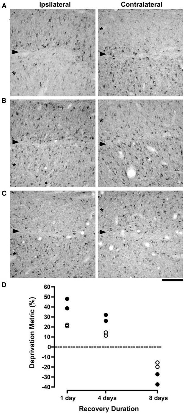
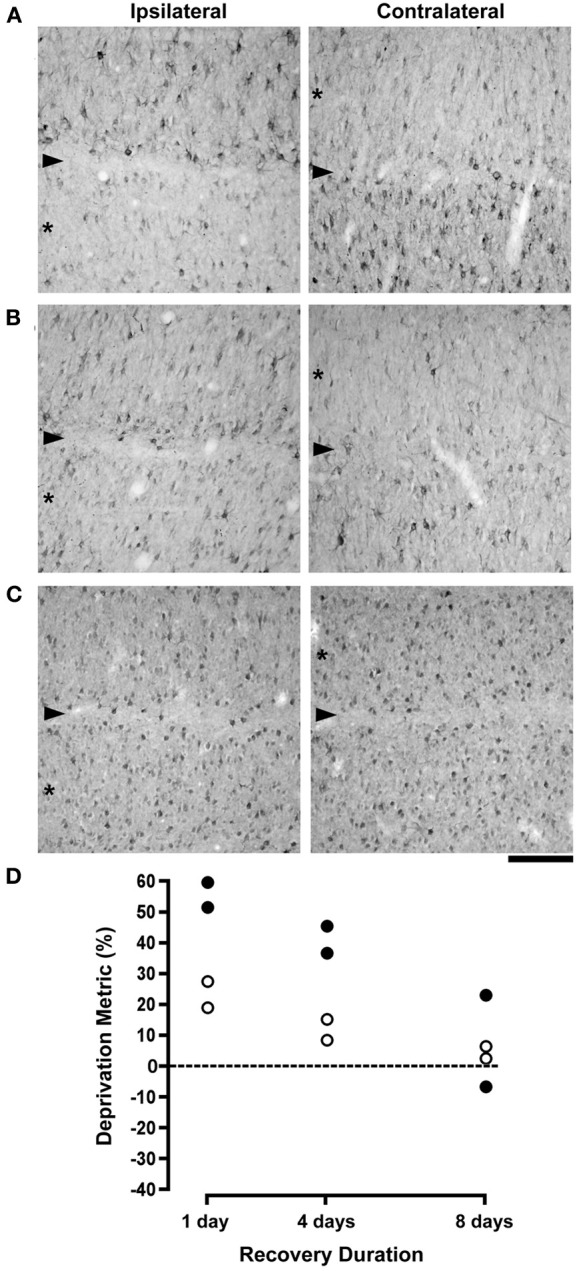
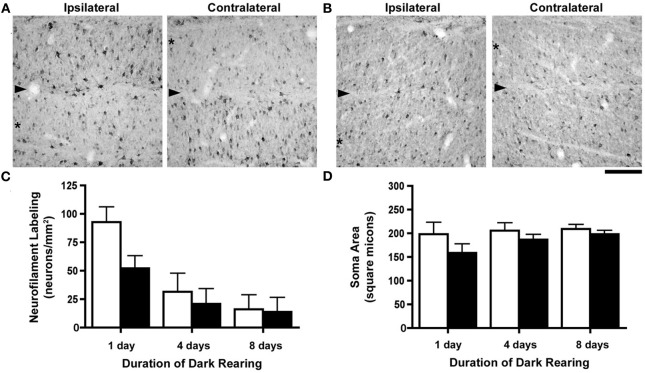
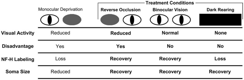
Postnatal development of the mammalian geniculostriate visual pathway is partly guided by visually driven activity. Disruption of normal visual input during certain critical periods can alter the structure of neurons, as well as their connections and functional properties. Within the layers of the dorsal lateral geniculate nucleus (dLGN), a brief early period of monocular deprivation can alter the structure and soma size of neurons within deprived-eye-receiving layers. This modification of structure is accompanied by a marked reduction in labeling for neurofilament protein, a principle component of the stable cytoskeleton. This study examined the extent of neurofilament recovery in monocularly deprived cats that either had their deprived eye opened (binocular recovery), or had the deprivation reversed to the fellow eye (reverse occlusion). The loss of neurofilament and the reduction of soma size caused by monocular deprivation were ameliorated equally and substantially in both recovery conditions after 8 days. The degree to which this recovery was dependent on visually driven activity was examined by placing monocularly deprived animals in complete darkness. Though monocularly deprived animals placed in darkness showed recovery of soma size in deprived layers, the manipulation catalyzed a loss of neurofilament labeling that extended to non-deprived layers as well. Overall, these results indicate that both recovery of soma size and neurofilament labeling is achieved by removal of the competitive disadvantage of the deprived eye. However, while the former occurred even in the absence of visually driven activity, recovery of neurofilament did not. The finding that a period of darkness produced an overall loss of neurofilament throughout the dLGN suggests that this experiential manipulation may cause the visual pathways to revert to an earlier more plastic developmental stage. It is possible that short periods of darkness could be incorporated as a component of therapeutic measures for treatment of deprivation-induced disorders such as amblyopia.

Keywords: cytoskeleton; dark-rearing; lateral geniculate nucleus; monocular deprivation; neurofilament; plasticity; recovery; reverse occlusion.


Figures

Figure 1
Figure 1
The effect of different durations of RO on NF-H labeling and on neuron size in the A laminae of the dLGN. Photomicrographs in (A–C) show NF-H labeling in the left and right dLGN (respectively, ipsilateral and contralateral to the deprived eye) where the originally deprived layer in each image is indicated by an asterisk, and the boundary between A layers is identified with an arrowhead. The effect of a preceding week-long period of monocular deprivation was still clearly evident in the originally deprived layers of the dLGN after RO for 1 day (A). Following 4 days of RO there was a recovery of NF-H labeling in originally deprived layers, but the originally non-deprived layers continued to show normal-looking NF-H labeling (B). By 8 days of RO there was a complete reversal of the deprivation effect so that originally deprived layers showed normal levels of NF-H labeling while labeling in the originally non-deprived layers was substantially reduced (C). Assessment of the recovery of neurofilament-positive cell density and neuron size in the dLGN was assessed with a deprivation metric (see Methods) that revealed a marked reversal of both NF-H labeling and neuron size. The size of neurons in originally deprived layers, which were smaller than originally non-deprived neurons at 1 day of RO, were larger than originally non-deprived neurons by almost the same amount following 8 days of RO (open circles in D). Likewise, originally deprived layers of the dLGN that exhibited comparatively fewer NF-H immunopositive neurons after 1 day of RO, contained considerably more NF-H labeled neurons following 8 days of RO (solid circles in D). Therefore, RO for 8 days fully reversed the effect of monocular deprivation on neuron size and NF-H labeling. The dashed line in (D) represents the point at which no difference exists between measurements from right and left eye layers. Scale bar = 100 μm.
Figure 2
Figure 2
The effects of different durations of binocular recovery following monocular deprivation are shown. Images in (A–C) show NF-H labeling in the (A) laminae of the left (ispilateral to the deprived eye) and right (contralateral to the deprived eye) dLGN, with originally deprived layers indicated by asterisks, and the boundary between layers identified with an arrowhead. Labeling for NF-H remained substantially reduced in deprived layers after providing binocular vision for 1 day, indicating that there was little if any recovery with this small duration of binocular exposure (A). Subsequent to 4 days of binocular vision, the effect of monocular deprivation was still evident but noticeably reduced, as deprived layers showed the beginning of a recovery of NF-H labeling (B). Labeling for NF-H in deprived layers appeared fully normal and indistinguishable from non-deprived layers after 8 days of binocular recovery (C). Results from the deprivation metric are plotted in (D), which reveals a gradual recovery of neuron size and NF-H labeling that was virtually complete following 8 days of binocular recovery. The dashed line in (D) represents the point at which no difference exists between measurements from right and left eye layers. Scale bar = 100 μm.
Figure 3
Figure 3
The consequence of different durations of dark-rearing following a week-long period of monocular deprivation. Images of the left (ipsilateral to the deprived eye) and right (contralateral to the deprived eye) dLGN after 1 day of dark-rearing (A) demonstrate a strong monocular deprivation effect with reduced NF-H labeling in originally deprived layers (asterisks) and strong labeling in originally non-deprived layers. The boundary between dLGN layers is indicated by an arrowhead. Following 8 days of dark-rearing we observed a reduction of labeling for NF-H in both originally deprived layers as well as layers that had not been deprived (B). We have graphed our measurements of NF-H labeling and neuron size (Mean + SD) in separate graphs because the deprivation metric is not ideal due to the extremely low densities of immunopositive neurons with extended dark-rearing. Density measurements of NF-H labeling were congruent with our observations from tissue, namely that there was a reduction in labeling after dark-rearing for 4 days, and a further reduction after 8 days (C). By 8 days of dark-rearing the effect of monocular deprivation was no longer evident because of the overall reduction of labeling in originally deprived and non-deprived layers. The difference in neuron size produced by monocular deprivation, still evident after a single day of dark-rearing, was reduced after 4 days, and even further so after 8 days of dark-rearing (D). Scale bar = 100 μm.
Figure 4
Figure 4
Schematic of the results from our investigation of NF-H labeling and neuron soma size in the dLGN across three recovery regimes (bolded text) following a period of early monocular deprivation. These results are presented alongside those from monocular lid suture from previous studies (Wiesel and Hubel, ; Guillery, ; Kutcher and Duffy, 2007). Comparison of the effect of NF-H labeling and soma size after monocular and binocular deprivation by lid suture suggests that the loss of NF-H and reduction in neuron size following monocular deprivation are the consequence of deprived neurons being put at a competitive disadvantage, and not because of a reduction in visually driven activity, which similarly occurs with binocular deprivation but without a reduction in NF-H NF-H (Kutcher and Duffy, 2007). Recovery of NF-H after monocular deprivation is achieved when the disadvantaged state of the deprived eye is relieved either by reverse occlusion or by restoration of binocular vision. It is noteworthy that the deprived eye need not be placed at a competitive advantage for this recovery to occur. Recovery of NF-H is evidently dependent upon visually driven activity because dark-rearing blocks recovery and leads to a pronounced overall loss of labeling. We, therefore, conclude that recovery of NF-H requires removal of the competitive disadvantage as well as visually driven activity. The recovery of neuron soma size is also achieved when the competitive disadvantage of the deprived eye is relieved, either by reverse occlusion or provision of binocular vision, and unlike recovery of NF-H, the recovery of neuron size is evidently not dependent on visually driven activity as deprived neurons recover in complete darkness.

Similar articles

Cited by

References

    1. Antonini A., Stryker M. P. (1993). Rapid remodeling of axonal arbors in the visual cortex. Science 260, 1819–1821. 10.1126/science.8511592 - DOI - PubMed
    1. Antonini A., Stryker M. P. (1998). Effect of sensory disuse on geniculate afferents to cat visual cortex. Vis. Neurosci. 15, 401–409. - PMC - PubMed
    1. Barry D. M., Millecamps S., Julien J.-P., Garcia M. L. (2007). New movements in neurofilament transport, turnover and disease. Exp. Cell Res. 313, 2110–2120. 10.1016/j.yexcr.2007.03.011 - DOI - PubMed
    1. Beaver C. J., Mitchell D. E., Robertson H. A. (1993). Immunohistochemical study of the patter of rapid expression of C-Fos protein in the visual cortex of dark-reared kittens following initial exposure to light. J. Comp. Neurol. 333, 469–484. 10.1002/cne.903330402 - DOI - PubMed
    1. Bickford M. E., Guido W., Godwin D. W. (1998). Neurofilament proteins in Y-cells of the cat lateral geniculate nucleus: normal expression and alteration with visual deprivation. J. Neurosci. 18, 6549–6557. - PMC - PubMed