

White matter atrophy and cognitive dysfunctions in neuromyelitis optica
- PMID: 22509264
- PMCID: PMC3317931
- DOI: 10.1371/journal.pone.0033878
White matter atrophy and cognitive dysfunctions in neuromyelitis optica
Abstract
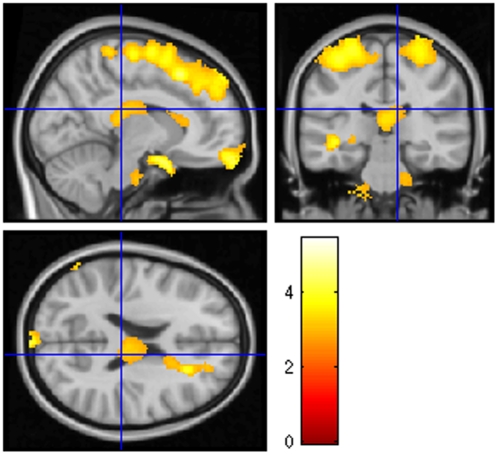
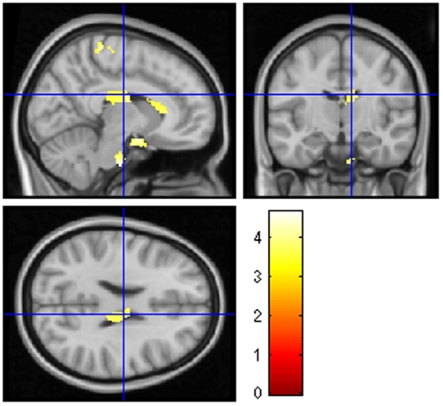
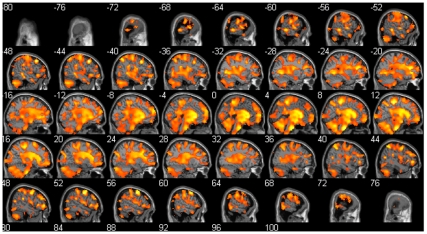
Neuromyelitis optica (NMO) is an inflammatory disease of central nervous system characterized by optic neuritis and longitudinally extensive acute transverse myelitis. NMO patients have cognitive dysfunctions but other clinical symptoms of brain origin are rare. In the present study, we aimed to investigate cognitive functions and brain volume in NMO. The study population consisted of 28 patients with NMO and 28 healthy control subjects matched for age, sex and educational level. We applied a French translation of the Brief Repeatable Battery (BRB-N) to the NMO patients. Using SIENAx for global brain volume (Grey Matter, GM; White Matter, WM; and whole brain) and VBM for focal brain volume (GM and WM), NMO patients and controls were compared. Voxel-level correlations between diminished brain concentration and cognitive performance for each tests were performed. Focal and global brain volume of NMO patients with and without cognitive impairment were also compared. Fifteen NMO patients (54%) had cognitive impairment with memory, executive function, attention and speed of information processing deficits. Global and focal brain atrophy of WM but not Grey Matter (GM) was found in the NMO patients group. The focal WM atrophy included the optic chiasm, pons, cerebellum, the corpus callosum and parts of the frontal, temporal and parietal lobes, including superior longitudinal fascicle. Visual memory, verbal memory, speed of information processing, short-term memory and executive functions were correlated to focal WM volumes. The comparison of patients with, to patients without cognitive impairment showed a clear decrease of global and focal WM, including brainstem, corticospinal tracts, corpus callosum but also superior and inferior longitudinal fascicles. Cognitive impairment in NMO patients is correlated to the decreased of global and focal WM volume of the brain. Further studies are needed to better understand the precise origin of cognitive impairment in NMO patients, particularly in the WM.
Conflict of interest statement
Figures
References
-
- Gault F. De la Neuromyélite Optique aiguë. Thèse à la faculté de Médecine et de Pharmacie de Lyon. 1894;981
-
- Lennon VA, Wingerchuk DM, Kryzer TJ, Pittock SJ, Lucchinetti CF, et al. A serum autoantibody marker of neuromyelitis optica: distinction from multiple sclerosis. Lancet. 2004;364:2106–2112. - PubMed
-
- Misu T, Fujihara K, Kakita A, Konno H, Nakamura M, et al. Loss of aquaporin 4 in lesions of neuromyelitis optica: distinction from multiple sclerosis. Brain. 2007;130:1224–1234. - PubMed
-
- Pittock SJ, Lennon VA, Krecke K, Wingerchuk DM, Lucchinetti CF, et al. Brain abnormalities in neuromyelitis optica. Arch Neurol. 2006;63:390–396. - PubMed
-
- Pittock SJ, Weinshenker BG, Lucchinetti CF, Wingerchuk DM, Corboy JR, et al. Neuromyelitis optica brain lesions localized at sites of high aquaporin 4 expression. Arch Neurol. 2006;63:964–968. - PubMed
MeSH terms
LinkOut - more resources
Full Text Sources
