. 2012;7(4):e34206.
doi: 10.1371/journal.pone.0034206. Epub 2012 Apr 3.

Unique cell type-specific junctional complexes in vascular endothelium of human and rat liver sinusoids

Affiliations

Unique cell type-specific junctional complexes in vascular endothelium of human and rat liver sinusoids

Cyrill Géraud et al. PLoS One. 2012.

Abstract

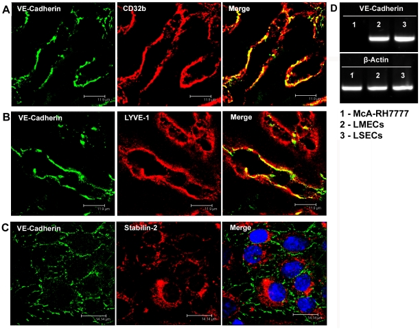
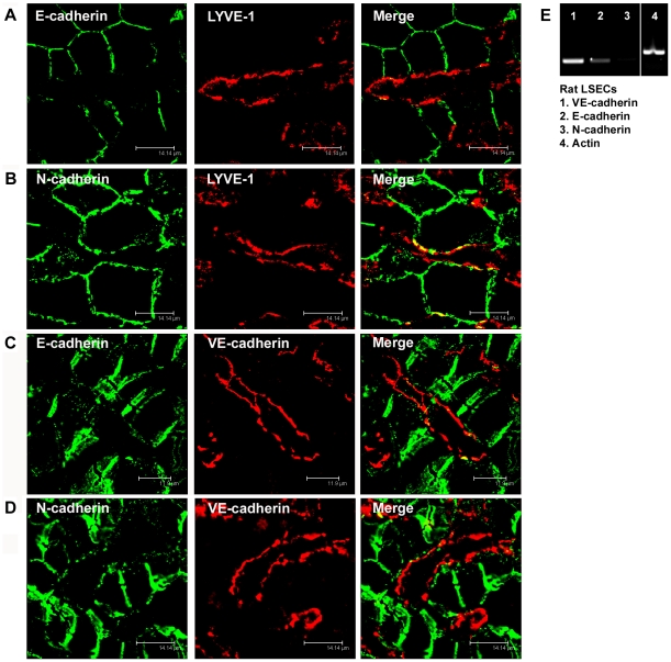
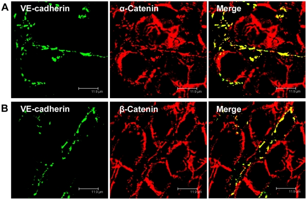
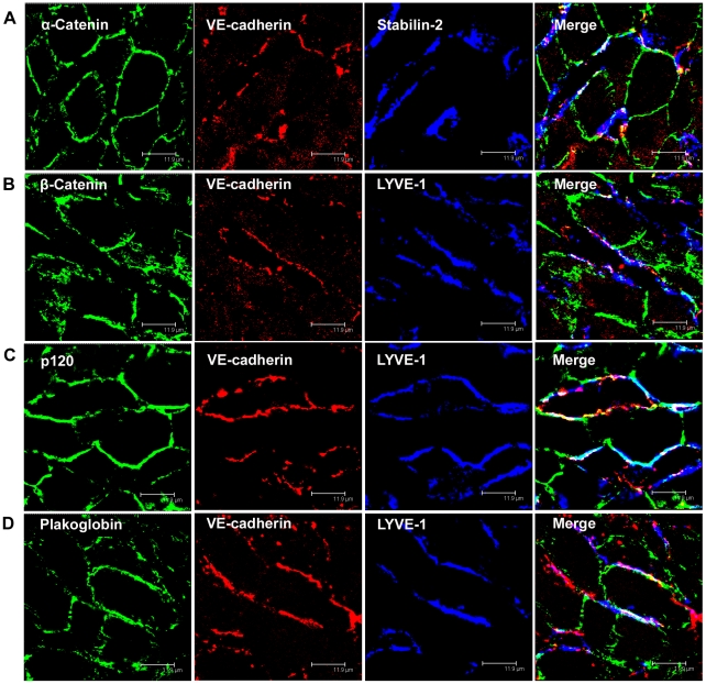
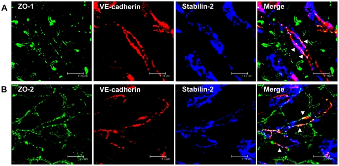
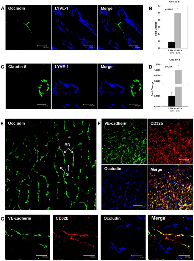
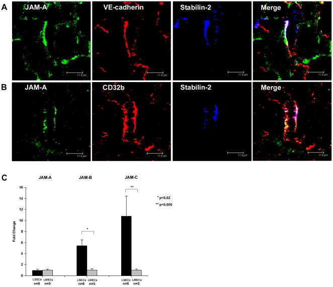
Liver sinusoidal endothelium is strategically positioned to control access of fluids, macromolecules and cells to the liver parenchyma and to serve clearance functions upstream of the hepatocytes. While clearance of macromolecular debris from the peripheral blood is performed by liver sinusoidal endothelial cells (LSECs) using a delicate endocytic receptor system featuring stabilin-1 and -2, the mannose receptor and CD32b, vascular permeability and cell trafficking are controlled by transcellular pores, i.e. the fenestrae, and by intercellular junctional complexes. In contrast to blood vascular and lymphatic endothelial cells in other organs, the junctional complexes of LSECs have not yet been consistently characterized in molecular terms. In a comprehensive analysis, we here show that LSECs express the typical proteins found in endothelial adherens junctions (AJ), i.e. VE-cadherin as well as α-, β-, p120-catenin and plakoglobin. Tight junction (TJ) transmembrane proteins typical of endothelial cells, i.e. claudin-5 and occludin, were not expressed by rat LSECs while heterogenous immunreactivity for claudin-5 was detected in human LSECs. In contrast, junctional molecules preferentially associating with TJ such as JAM-A, B and C and zonula occludens proteins ZO-1 and ZO-2 were readily detected in LSECs. Remarkably, among the JAMs JAM-C was considerably over-expressed in LSECs as compared to lung microvascular endothelial cells. In conclusion, we show here that LSECs form a special kind of mixed-type intercellular junctions characterized by co-occurrence of endothelial AJ proteins, and of ZO-1 and -2, and JAMs. The distinct molecular architecture of the intercellular junctional complexes of LSECs corroborates previous ultrastructural findings and provides the molecular basis for further analyses of the endothelial barrier function of liver sinusoids under pathologic conditions ranging from hepatic inflammation to formation of liver metastasis.


Conflict of interest statement

Competing Interests: The authors have declared that no competing interests exist.

Figures

Figure 1
Figure 1. VE-cadherin is expressed in liver sinusoidal endothelial cells in rats and humans.
(A) Immunofluorescent co-staining of human liver cryosections with anti-VE-cadherin (green) and anti-CD32b (red) antibodies. (B) Immunofluorescent co-staining of rat liver cryosections with anti-VE-cadherin (green) and anti-LYVE-1 (red) antibodies. (C) Immunofluorescent co-staining of isolated rat LSECs with anti-VE-cadherin (green) and anti-Stabilin-2 (red) antibodies. Toto3 (blue) was used to counterstain the cell nuclei. Images were acquired using laser scanning confocal microscopy. Bars 11.9 µm (A, B), 14.14 µm (C). (D) Reverse transcriptase-PCR with mRNA isolated from rat hepatoma McA-RH7777 cell line (1), freshly isolated rat LMECs (2), and freshly isolated rat LSECs (3). Primers specific for VE-cadherin or β-actin were used.
Figure 2
Figure 2. E- and N-cadherin are absent in liver sinusoidal endothelial cells.
(A, B) Immunofluorescent co-staining of rat liver cryosections with anti-E-cadherin (A, green) or anti-N-cadherin (B, green) and anti-LYVE-1 (A, B, red) antibodies. (C, D) Immunofluorescent co-staining of human liver cryosections with anti-E-cadherin (C, green) or anti-N-cadherin (D, green) and anti-VE-cadherin (C, D, red) antibodies. Images were acquired using laser scanning confocal microscopy. Bars 14.14 µm (A, B, D), 11.9 µm (C). (E) Reverse transcriptase-PCR with mRNA of freshly isolated rat LSECs. Primers specific for VE-cadherin (1), E-cadherin (2), N-cadherin (3) or β-actin (4) were used.
Figure 3
Figure 3. α-Catenin and β-Catenin co-localize with VE-cadherin in human LSECs.
Immunofluorescent co-staining of human liver cryosections with anti-VE-cadherin (A, B, green), anti-α-Catenin (A, red), and anti-β-catenin (B, red) antibodies. Images were acquired using laser scanning confocal microscopy. Bars 11.9 µm.
Figure 4
Figure 4. α-catenin, β-catenin, p120-catenin, and plakoglobin co-localize with VE-cadherin in rat liver sinusoidal endothelial cells.
(A-D) Immunofluorescent co-staining of rat liver cryosections with anti-α-Catenin (A, green), anti-β-Catenin (B, green), anti-p120-Catenin (C, green), anti-Plakoglobin (D, green), anti-VE -cadherin (A-D, red), anti-Stabilin-2 (A, blue), and anti-LYVE-1 (B-D, blue) antibodies. Images were acquired using laser scanning confocal microscopy. Bars 11.9 µm.
Figure 5
Figure 5. ZO-1 and ZO-2 localize to VE-cadherin-containing cell-cell junctions in rat liver sinusoidal endothelial cells.
(A, B) Immunofluorescent co-staining of rat liver cryosections with anti-ZO-1 (A, green), anti-ZO-2 (B, green), anti-VE-cadherin (A, B, red), and anti-Stabilin-2 (A, B, blue) antibodies. Images were acquired using laser scanning confocal microscopy. Bars 11.9 µm.
Figure 6
Figure 6. Immunoflurescent and qRT-PCR analysis of occludin and claudin-5 expression in liver sinusoidal endothelial cells.
(A, C) Immunofluorescent co-staining of rat liver cryosections with anti-Occludin (A, green), anti-Claudin-5 (C, green), and anti-LYVE-1 (A, C, blue) antibodies. (E) Immunofluorescent staining of a liver sample obtained from the patient 2 with anti-Occludin (green) antibody; BD – bile ducts, S – liver sinusoids. (F, G) Immunofluorescent co-staining of liver samples obtained from the patients 6 (F) and 4 (G) with anti-VE-cadherin (F, G, green), anti-CD32b (F, G, red), and anti-Occludin (F, G, blue) antibodies. Images were acquired using laser scanning confocal microscopy. Bars 11.9 µm (A, C), 47.62 µm (E, F), 14.14 µm (G). (B, D) Quantitative reverse transcriptase-PCR with mRNA isolated from rat LSECs and rat LMECs (n indicates the number of samples analyzed, error bars represent SEM). Primers specific for Occludin (B), Claudin-5 (D), and β-Actin as normalizer were used.
Figure 7
Figure 7. Expression of JAM-family members in liver sinusoidal endothelial cells.
(A) Immunofluorescent co-staining of rat liver cryosections with anti-JAM-A (green), anti-VE-cadherin (red), and anti-Stabilin-2 (blue) antibodies. (B) Immunofluorescent co-staining of human liver cryosections with anti-JAM-A (green), anti-CD32b (red), and anti-Stabilin-2 (blue) antibodies. Images were acquired using laser scanning confocal microscopy. Bars 11.9 µm. (C) Quantitative reverse transcriptase-PCR with mRNA isolated from rat LSECs and rat LMECs (n indicates the number of samples analyzed, error bars represent SEM). Primers specific for JAM-A, JAM-B, JAM-C, and β-Actin as normalizer were used.

References

    1. Perri R, Shah V. Chapter 5: Hepatic Sinusoidal Endothelial Cells. In: Dufour JF, Clavien PA, editors. Signaling pathways in liver diseases. Berlin Heidelberg: Springer-Verlag; 2005. pp. 53–62.
    1. Si-Tayeb K, Lemaigre FP, Duncan SA. Organogenesis and development of the liver. Dev Cell. 2010;18:175–189. - PubMed
    1. Aird WC. Phenotypic heterogeneity of the endothelium: II. Representative vascular beds. Circ Res. 2007;100:174–190. - PubMed
    1. Steinhoff G, Behrend M, Schrader B, Duijvestijn AM, Wonigeit K. Expression patterns of leukocyte adhesion ligand molecules on human liver endothelia. Lack of ELAM-1 and CD62 inducibility on sinusoidal endothelia and distinct distribution of VCAM-1, ICAM-1, ICAM-2, and LFA-3. Am J Pathol. 1993;142:481–488. - PMC - PubMed
    1. Lalor PF, Adams DH. Adhesion of lymphocytes to hepatic endothelium. Mol Pathol. 1999;52:214–219. - PMC - PubMed

Publication types

MeSH terms