. 2012;7(4):e34250.
doi: 10.1371/journal.pone.0034250. Epub 2012 Apr 3.

Phosphorylation of Ubc9 by Cdk1 enhances SUMOylation activity

Affiliations

Phosphorylation of Ubc9 by Cdk1 enhances SUMOylation activity

Yee-Fun Su et al. PLoS One. 2012.

Abstract

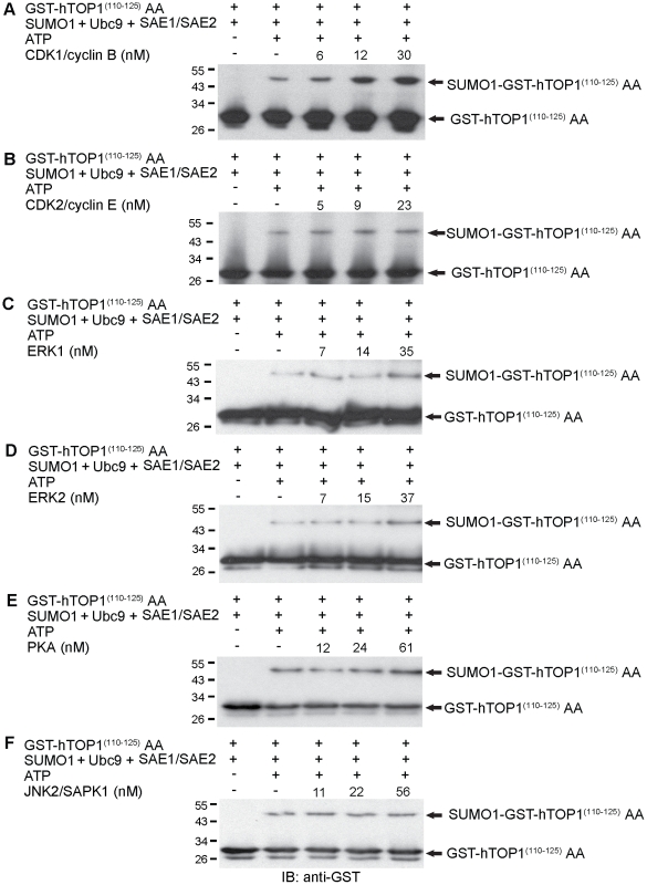
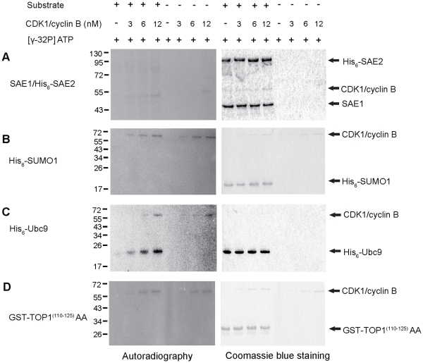
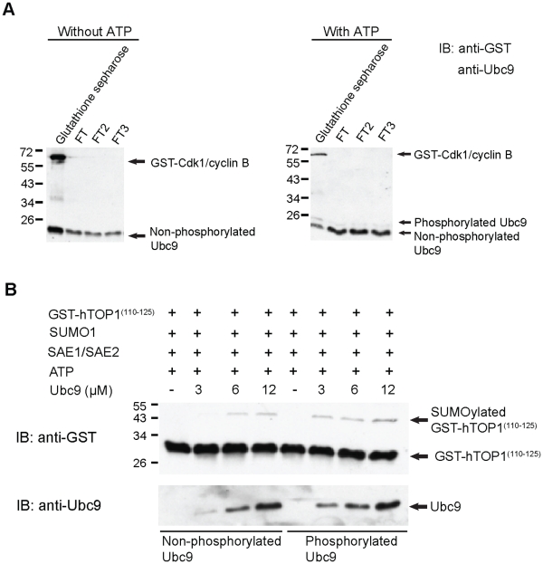
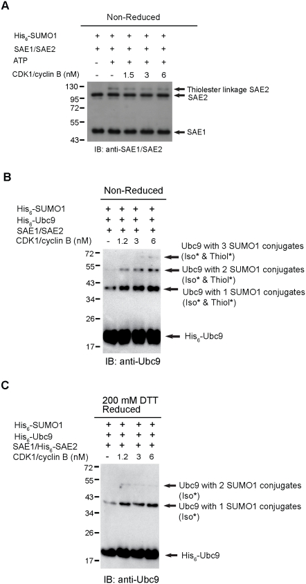
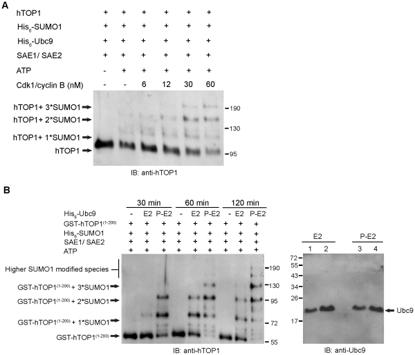
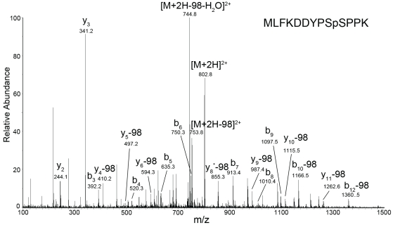
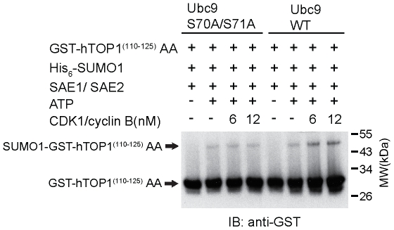
Increasing evidence has pointed to an important role of SUMOylation in cell cycle regulation, especially for M phase. In the current studies, we have obtained evidence through in vitro studies that the master M phase regulator CDK1/cyclin B kinase phosphorylates the SUMOylation machinery component Ubc9, leading to its enhanced SUMOylation activity. First, we show that CDK1/cyclin B, but not many other cell cycle kinases such as CDK2/cyclin E, ERK1, ERK2, PKA and JNK2/SAPK1, specifically enhances SUMOylation activity. Second, CDK1/cyclin B phosphorylates the SUMOylation machinery component Ubc9, but not SAE1/SAE2 or SUMO1. Third, CDK1/cyclin B-phosphorylated Ubc9 exhibits increased SUMOylation activity and elevated accumulation of the Ubc9-SUMO1 thioester conjugate. Fourth, CDK1/cyclin B enhances SUMOylation activity through phosphorylation of Ubc9 at serine 71. These studies demonstrate for the first time that the cell cycle-specific kinase CDK1/cyclin B phosphorylates a SUMOylation machinery component to increase its overall SUMOylation activity, suggesting that SUMOylation is part of the cell cycle program orchestrated by CDK1 through Ubc9.


Conflict of interest statement

Competing Interests: The authors have declared that no competing interests exist.

Figures

Figure 1
Figure 1. CDK1/cyclin B stimulates SUMOylation in vitro.
(A-F) Various concentrations of different recombinant kinases, CDK1/cyclin B (6 nM, 12 nM and 30 nM), CDK2/cyclin E (5 nM, 9 nM and 23 nM), ERK1 (7 nM, 14 nM and 35 nM), ERK2 (7 nM, 15 nM and 37 nM), PKA (12 nM, 24 nM and 61 nM) and JNK2/SAPK1 (11 nM, 22 nM and 56 nM), were added to in vitro SUMOylation reaction mixtures containing His6-SUMO1, His6-Ubc9, SAE1/His6-SAE2, GST-hTOP1 (110–125) AA and ATP at 37°C for 60 min. The reactions were then boiled in SDS sample buffer and analyzed by 15% SDS-PAGE, followed by immunoblotting with anti-GST antibody.
Figure 2
Figure 2. CDK1/cyclin B phosphorylates Ubc9 in vitro.
(A) In vitro phosphorylation of SAE1/SAE2 by CDK1/cyclin B. CDK1/cyclin B (3 nM, 6 nM and 12 nM) was incubated with (left) or without (right) SAE1/His6-SAE2 for 30 min at 30°C in the presence of [γ-32P] ATP. The reactions were analyzed by 10% SDS-PAGE followed by Coomassie blue staining and autoradiography. (B) In vitro phosphorylation of His6-SUMO1 by CDK1/cyclin B. Various concentrations of CDK1/cyclin B (as mentioned above) were incubated with (left) or without His6-SUMO1 (right) in the presence of [γ-32P] ATP for 30 min at 30°C. (C) In vitro phosphorylation of Ubc9 by CDK1/cyclin B. Various concentrations of CDK1/cyclin B (as mentioned above) were incubated with (left panel) or without His6-Ubc9 (right panel) in the presence of [γ-32P] ATP for 30 min at 30°C. (D) In vitro phosphorylation of GST-hTOP1 (110–125)AA. Various concentrations of CDK1/cyclin B (as mentioned above) were incubated with (left) or without GST-hTOP1(110–125)AA (right) in the presence of [γ-32P] ATP. Reaction mixtures of B, C and D were analyzed by 15% SDS-PAGE followed by Coomassie blue staining and autoradiography.
Figure 3
Figure 3. CDK1/cyclin B-phosphorylated Ubc9 exhibits elevated SUMOylation activity.
(A) Purified His6-Ubc9 was incubated with GST-CDK1/cyclin B in the presence or absence of ATP for 30 min at 30°C. The GST-CDK1/cyclin B was then removed by passing through glutathione sepharose. The bound fraction of glutathione sepharose (contains phosphorylated Ubc9) and flow through fractions (FT, FT2, FT3) were analyzed by 12.5% SDS-PAGE, followed by immunoblotting with anti-GST and anti-Ubc9 antibody. (B) 3 µM, 6 µM and 12 µM of phosphorylated Ubc9 (flow through fraction 3) and non-phosphorylated Ubc9 (flow through fraction 3) were individually incubated with His6-SUMO1, SAE1/His6-SAE2, GST-hTOP1(110–125) and ATP for 60 min at 37°C. The SUMOylation reaction mixtures were then analyzed by 15% SDS-PAGE, followed by immunoblotting with anti-GST antibody. The amount of phosphorylated and non-phosphorylated Ubc9 for SUMOylation assay were detected by immunoblotting with anti-Ubc9 antibody.
Figure 4
Figure 4. CDK1/cyclin B upregulates SUMOylation by increasing thioester bond formation between phosphorylated Ubc9 and SUMO1 but not between SAE1/SAE2 and SUMO1.
(A) To examine the thioester bond formation between His6-SUMO1 and SAE1/His6-SAE2, purified His6-SUMO1 and SAE1/His6-SAE2 were incubated with various concentrations of CDK1/cyclin B (1.5 nM, 3 nM and 6 nM) in the presence or absence of ATP. Reaction mixtures were run on a 10% SDS-PAGE and immunoblotted with anti-SAE1/SAE2 antibody. (B) Ubc9 thioester conjugation assay was performed by incubating purified His6-Ubc9, SAE1/His6-SAE2 and His6-SUMO1 with various concentrations of CDK1/cyclin B (1.2 nM, 3 nM and 6 nM) in the presence of ATP for 60 min at 37°C. Samples were then resolved on a 15% SDS-PAGE and probed with anti-Ubc9 antibody. (C) Ubc9 thioester conjugation assay in was performed similar as in (B), but at the end of the reaction, 200 mM of DTT was added to samples for overnight at 40°C. The reduced samples were run on a 15% SDS-PAGE, followed by immunoblotting with anti-Ubc9 antibody. Iso, isopeptide bond; Thiol, thioester bond.
Figure 5
Figure 5. CDK1/cyclin B enhances SUMOylation level of human TOP1 containing multiple SUMO conjugation site.
(A) Full-length protein of human topoisomerase I was incubated with His6-SUMO1, His6-Ubc9, SAE1/His-SAE2 and ATP in the presence of 6, 12, 30 and 60 nM of CDK1/cyclin B. The reaction was resolved on a 6% SDS-PAGE and immunoblotted with anti-topoisomerase I antibody. (B) Equal amount of CDK1/cyclin B phosphorylated Ubc9 (P-E2) and Ubc9 (E2) (6 µM at the final concentration) was individually incubated with His6-SUMO1, SAE1/His6-SAE2, GST-hTOP1(1–200) and ATP at 37°C for 30 min, 60 min and 120 min. The resulting SUMOylation reaction mixtures were run on a 8% SDS-PAGE, followed by immunoblotting with anti-topoisomerase I antibody. The amount of phosphorylated Ubc9 (P-E2) and Ubc9 (E2) for SUMOylation assay in Fig. 5B was determined by immunoblotting with anti-Ubc9 antibody. Lane 1 and 3 (right panel) were loaded with 1 µg Ubc9 and phosphorylated Ubc9, respectively. Lane 2 and lane 4 (right panel) were loaded with 2 µg of Ubc9 and phosphorylated Ubc9, respectively.
Figure 6
Figure 6. CDK1/cyclin B mediates phopshorylation of Ubc9 at Serine 71.
IMAC enriched tryptic peptides of Ubc9 were detected by CID tandem mass spectra. Precursor ion m/z 802.8 (charge state +2) representing the phosphopeptide MLFKDDYPSpSPPK. Residues bearing phosphate moieties are indicated with p. “b” and “y” ions series represent fragment ions containing the N- and C-termini of the peptide, respectively. Product ions marked with * resulted from elimination of NH3. The mass of 98 on the peaks was derived from neutral losses (-97.9769 Da) of phosphoric acid (H3PO4).
Figure 7
Figure 7. CDK1/cyclin B enhances SUMOylation activity through wild type Ubc9 but not mutant Ubc9 S70A/S71A.
Same amount of wild-type and mutant (S70A/S71A) of Ubc9 (at a concentration of 6 µM) was individually added to in vitro SUMOylation reaction mixtures containing His6-SUMO1, SAE1/His6-SAE2, GST-hTOP1 (110–125) AA and ATP in the presence of CDK1/cyclin B (6 nM and 12 nM) at 37°C for 60 min. The reactions were then boiled in SDS sample buffer and analyzed by 15% SDS-PAGE, followed by immunoblotting with anti-GST antibody.

References

    1. Hay RT. SUMO: a history of modification. Mol Cell. 2005;18:1–12. - PubMed
    1. Azuma Y, Arnaoutov A, Dasso M. SUMO-2/3 regulates topoisomerase II in mitosis. J Cell Biol. 2003;163:477–487. - PMC - PubMed
    1. Fernandez-Miranda G, de Castro IP, Carmena M, Aguirre-Portoles C, Ruchaud S, et al. SUMOylation modulates the function of Aurora-B kinase. J Cell Sci 123 (Pt. 2010;16):2823–2833. - PMC - PubMed
    1. Seufert W, Futcher B, Jentsch S. Role of a ubiquitin-conjugating enzyme in degradation of S- and M-phase cyclins. Nature. 1995;373:78–81. - PubMed
    1. al-Khodairy F, Enoch T, Hagan IM, Carr AM. The Schizosaccharomyces pombe hus5 gene encodes a ubiquitin conjugating enzyme required for normal mitosis. J Cell Sci 108 ( Pt. 1995;2):475–486. - PubMed

Publication types

MeSH terms