. 2012;7(4):e34477.
doi: 10.1371/journal.pone.0034477. Epub 2012 Apr 3.

Erythropoietin receptor signaling is membrane raft dependent

Affiliations

Erythropoietin receptor signaling is membrane raft dependent

Kathy L McGraw et al. PLoS One. 2012.

Abstract

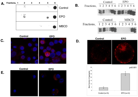
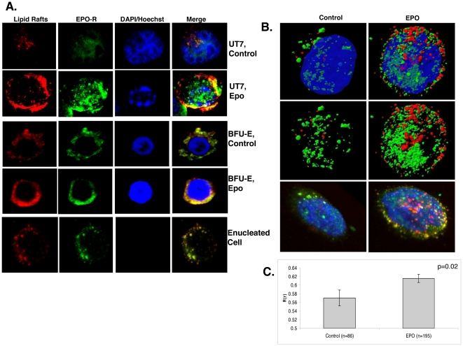
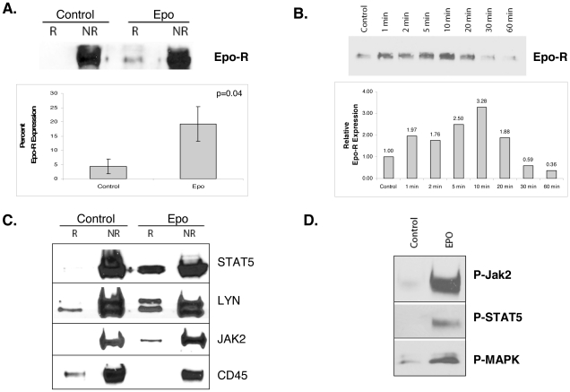
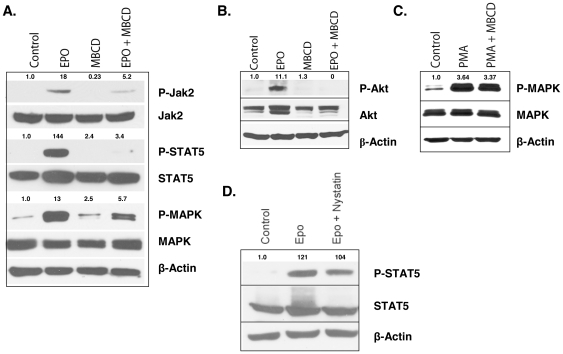
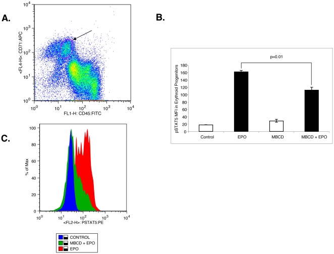
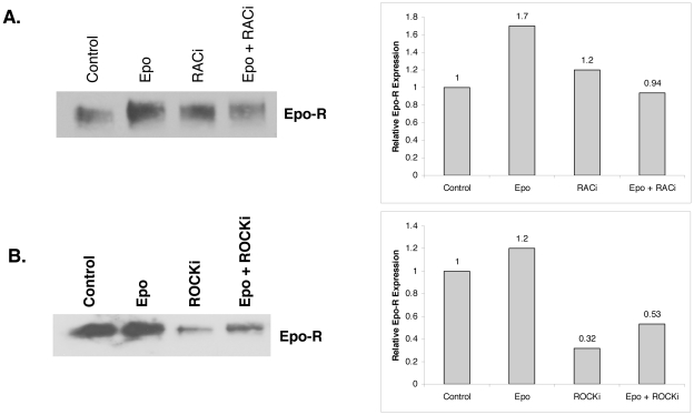
Upon erythropoietin (Epo) engagement, Epo-receptor (R) homodimerizes to activate JAK2 and Lyn, which phosphorylate STAT5. Although recent investigations have identified key negative regulators of Epo-R signaling, little is known about the role of membrane localization in controlling receptor signal fidelity. Here we show a critical role for membrane raft (MR) microdomains in creation of discrete signaling platforms essential for Epo-R signaling. Treatment of UT7 cells with Epo induced MR assembly and coalescence. Confocal microscopy showed that raft aggregates significantly increased after Epo stimulation (mean, 4.3±1.4(SE) vs. 25.6±3.2 aggregates/cell; p≤0.001), accompanied by a >3-fold increase in cluster size (p≤0.001). Raft fraction immunoblotting showed Epo-R translocation to MR after Epo stimulation and was confirmed by fluorescence microscopy in Epo stimulated UT7 cells and primary erythroid bursts. Receptor recruitment into MR was accompanied by incorporation of JAK2, Lyn, and STAT5 and their activated forms. Raft disruption by cholesterol depletion extinguished Epo induced Jak2, STAT5, Akt and MAPK phosphorylation in UT7 cells and erythroid progenitors. Furthermore, inhibition of the Rho GTPases Rac1 or RhoA blocked receptor recruitment into raft fractions, indicating a role for these GTPases in receptor trafficking. These data establish a critical role for MR in recruitment and assembly of Epo-R and signal intermediates into discrete membrane signaling units.


Conflict of interest statement

Competing Interests: The authors have declared that no competing interests exist.

Figures

Figure 1
Figure 1. Epo stimulation induces raft formation and aggregation.
(A) Dot blot detection of GM-1 in UT7 cell lysates in non-raft (fractions 5, 6) and raft fractions (fraction 2) with corresponding densitometry value in controls, and after Epo or MBCD treatment. Representative blot of at least three independent experiments. (B) Western immunoblot of Lyn in raft (R) (fractions 1–2) and non-raft (NR) fractions (fractions 4–6). Treatment with Epo increased Lyn kinase incorporation into raft fractions, whereas raft disruption by cholesterol depletion with MβCD precluded Lyn incorporation. Representative western of at least three independent experiments. (C) Immunofluorescence of UT7 cells showing an increase in raft (red) accumulation after Epo exposure. (D) Immunofluorescence of UT7 cells before and after Epo stimulation showing increased raft aggregates (red) in the plasma membrane and corresponding quantitation. (E) Immunofluorescence of primary erythroid bursts showing an increase in cellular membrane raft fluorescence intensity (red). Immunoflorescence experiments were repeated at least 3 times, representative micrographs displayed.
Figure 2
Figure 2. Epo-R co-localizes with lipid rafts.
(A) Confocal immunofluorescence of cells untreated or after Epo stimulation, lipid rafts:red, Epo-R:green, DAPI/Hoechst:blue. Right panel is a merged image showing lipid raft and Epo-R co-localization (yellow). UT7 cells are shown in rows 1 and 2, while human primary burst forming units are shown in rows 3 and 4, followed by a maturing, enucleated erythroid precursor in row 5. (B) Three dimensional rendering of UT7 cells either untreated (left) or after Epo treatment (right). Top two rows display isosurfacing of the rafts (red), Epo-R (green), and nucleus (Dapi, blue). Dapi was removed from the middle row to further visualize association of the receptor with rafts in the second row of panels. The bottom row displays volume rendering of the same cells to illustrate membrane colocalization (yellow). (C) Quantitation of colocalization in human primary erythroid cells. Values represent mean ± SE. Immunofluorescence experiments were repeated at least 3 times, representative micrographs provided.
Figure 3
Figure 3. Epo stimulation recruits signal effectors into raft fractions.
(A) Raft fractions (R) were separated from non-raft fractions (NR) and immunoblotted for Epo-R to investigate receptor translocation into rafts after Epo stimulation. Corresponding quantitation represents the mean ± SE of two independent experiments using four different Epo-R antibodies. (B) Raft fractions were isolated after stimulation with Epo at the indicated time points and immunoblotted for Epo-R. Results show that EpoR is recruited into rafts within 1 minute of Epo stimulation reaching maximum loading at 10 minutes, followed by gradual redistribution thereafter. Accompanying graphic quantitation of the representative experiment. (C) UT7 cells were starved overnight then treated with Epo for 10 min. After fractionation, the non-raft (NR) fractions and raft (R) fractions were pooled and immunoblotted for the indicated proteins. (D) Activated forms of Jak2, STAT5, and MAPK were also increased in the raft fractions after Epo stimulation. All westerns were repeated at least in duplicate.
Figure 4
Figure 4. Raft integrity is necessary for Epo-induced signaling.
(A) UT7 cells were starved for 2 h then pretreated with MBCD for 30 min and stimulated with 3 U/ml Epo for 10 min; lysates were immunoblotted with the indicated antibodies. (B) UT7/Epo cells were starved for 2 h then pretreated with MBCD for 30 min and stimulated with 3 U/ml Epo for 10 min. Lysates were immunoblotted with P-Akt. The findings show abrogation of Akt phosphorylation following MBCD pretreatment. (C) UT7 cells were pretreated with MBCD for 30 min, then stimulated with PMA for 30 min. (D) UT7 cells were starved for 2 h then pretreated with Nystatin for 30 min and stimulated with Epo for 10 min. Immunoblots for phospho-STAT5, STAT5, and β-actin antibodies with densitometry analysis. All westerns are representative of at least 2 independent experiments.
Figure 5
Figure 5. Cholesterol depletion attenuates Epo-induced STAT5 phosphorylation in primary erythroid progenitors.
(A) Bone marrow mononuclear cells from a normal donor were isolated then stained with CD71:APC, CD45:FITC, and P-STAT5:PE. CD71Hi/CD45dim cells representing erythroid progenitors were gated. (B) Graphic comparison of geometric mean florescence intensities, mean ± standard error from 3 independent experiments. (C) Representative flow histogram showing shift in phospho-STAT5 florescence intensity in primary erythroid progenitors treated with Epo with or without MβCD.
Figure 6
Figure 6. Recruitment of Epo-R into lipid rafts is dependent on Rac1 and RhoA GTPase activation.
(A) Raft fractions were isolated from UT7 cells pretreated with 100 nM Rac1 inhibitor for 1 hr prior to Epo stimulation then immunoblotted for Epo-R with corresponding quantitation. (B) Raft fractions were isolated from UT7 cells pretreated with 100 uM ROCK inhibitor (Y-27632) for 1 h prior to Epo stimulation then immunoblotted for Epo-R with corresponding densitometry analysis. Westerns are representative of two independent experiments.

References

    1. Witthuhn B, Quelle F, Silvennoinen O, Yi T, Tang B, et al. Jak2 associates with the erythropoietin receptor and is tyrosine phosphorylated and activated following stimulation with erythropoietin. Cell. 1993;74:227–236. - PubMed
    1. Wojchowski D, Gregory R, Miller C, Pandit A, Pircher T. Signal transduction in the erythropoietin receptor system. Experimental cell research. 1999;253:143–156. - PubMed
    1. Rane SG, Reddy EP. JAKs, STATs and Src kinases in hematopoiesis. Oncogene. 2002;21:3334–3358. - PubMed
    1. Karur VG, Lowell CA, Besmer P, Agosti V, Wojchowski DM. Lyn kinase promotes erythroblast expansion and late-stage development. Blood. 2006;108:1524–1532. - PMC - PubMed
    1. Tilbrook PA, Ingley E, Williams JH, Hibbs ML, Klinken SP. Lyn tyrosine kinase is essential for erythropoietin-induced differentiation of J2E erythroid cells. The EMBO journal. 1997;16:1610–1619. - PMC - PubMed

Publication types

MeSH terms