. 2012;7(4):e34600.
doi: 10.1371/journal.pone.0034600. Epub 2012 Apr 3.

Estrogen prevents oxidative damage to the mitochondria in Friedreich's ataxia skin fibroblasts

Affiliations

Estrogen prevents oxidative damage to the mitochondria in Friedreich's ataxia skin fibroblasts

Timothy E Richardson et al. PLoS One. 2012.

Abstract

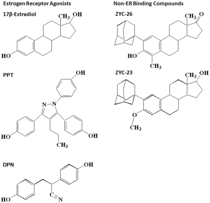
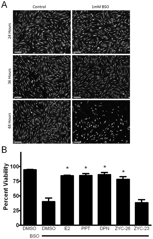
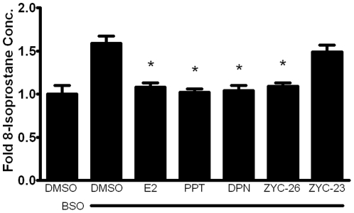
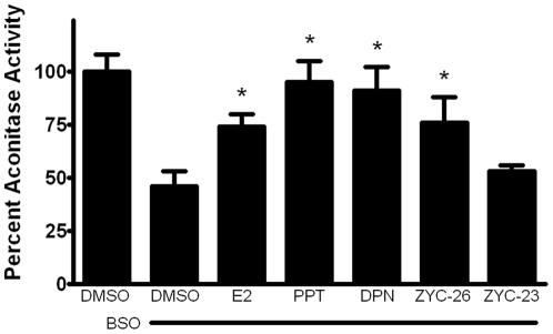
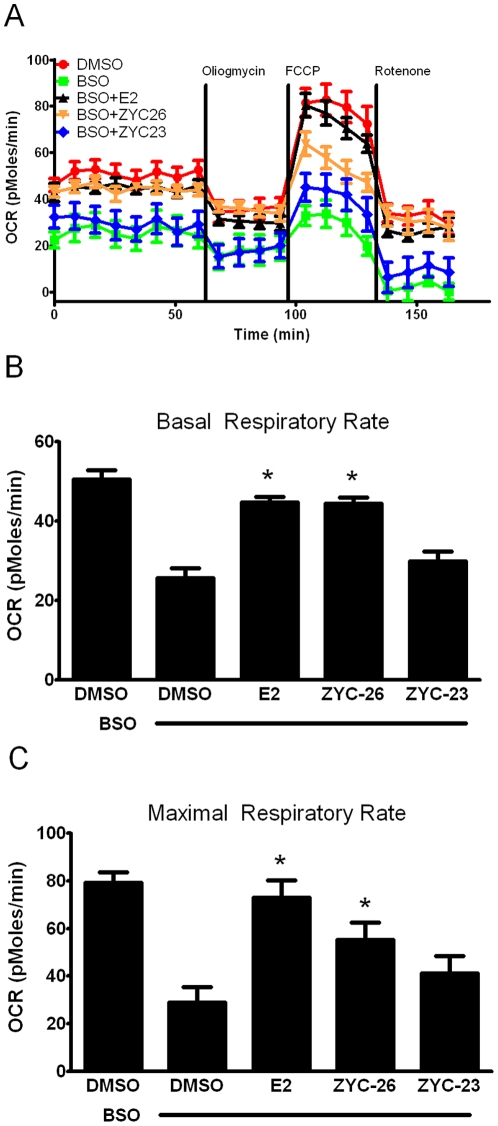
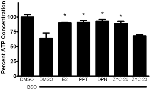
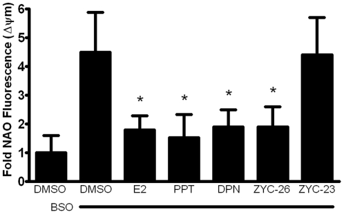
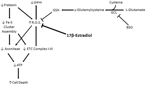
Estrogen and estrogen-related compounds have been shown to have very potent cytoprotective properties in a wide range of disease models, including an in vitro model of Friedreich's ataxia (FRDA). This study describes a potential estrogen receptor (ER)-independent mechanism by which estrogens act to protect human FRDA skin fibroblasts from a BSO-induced oxidative insult resulting from inhibition of de novo glutathione (GSH) synthesis. We demonstrate that phenolic estrogens, independent of any known ER, are able to prevent lipid peroxidation and mitochondrial membrane potential (ΔΨm) collapse, maintain ATP at near control levels, increase oxidative phosphorylation and maintain activity of aconitase. Estrogens did not, however, prevent BSO from depleting GSH or induce an increased expression level of GSH. The cytoprotective effects of estrogen appear to be due to a direct overall reduction in oxidative damage to the mitochondria, enabling the FRDA fibroblast mitochondria to generate sufficient ATP for energy requirements and better survive oxidative stress. These data support the hypothesis that phenol ring containing estrogens are possible candidate drugs for the delay and/or prevention of FRDA symptoms.


Conflict of interest statement

Competing Interests: The authors have declared that no competing interests exist.

Figures

Figure 1
Figure 1. Structures of compounds assessed for protection against BSO toxicity in FRDA fibroblasts.
Figure 2
Figure 2. Western blot showing the presence of small amounts of ERβ and the absence of ERα in FRDA fibroblasts compared with 661W photoreceptor cells.
Figure 3
Figure 3
A.) Calcein AM imaging demonstrating cell viability between vehicle control and BSO treatment groups at 24, 36 and 48 hours. Scale bar = 200 µm. B.) Effects of E2, PPT, DPN, ZYC-26 and ZYC-23 on cell viability in BSO-treated FRDA fibroblasts. All steroid concentrations were 100 nM, DMSO concentration was 0.1% and BSO concentration was 1 mM. Depicted are mean ± SD for n = 8 per group. * indicated p<0.05 versus BSO alone-treated cells.
Figure 4
Figure 4. Effects of E2, PPT, DPN, ZYC-26 and ZYC-23 on intracellular lipid peroxidation in BSO-treated FRDA fibroblasts.
All steroid concentrations were 100 nM, DMSO concentration was 0.1% and BSO concentration was 1 mM. Depicted are mean ± SD for n = 8 per group. * indicated p<0.05 versus BSO alone-treated cells. 1.0 normalized 8-isoprostane control concentration = 6.23 pg/mL.
Figure 5
Figure 5. Effects of E2, PPT, DPN, ZYC-26 and ZYC-23 on the activity of aconitase in BSO-treated FRDA fibroblasts.
All steroid concentrations were 100 nM, DMSO concentration was 0.1% and BSO concentration was 1 mM. Depicted are mean ± SD for n = 8 per group. * indicated p<0.05 versus BSO alone-treated cells.
Figure 6
Figure 6. Effects of E2 and ZYC-26 on mitochondrial function in BSO-treated FRDA fibroblasts.
A.) Oxygen consumption rate (OCR; in pMoles/min) B.) Basal respiratory rate (in pMoles/min) C.) Maximal respiratory rate (in pMoles/min) All steroid concentrations were 100 nM, DMSO concentration was 0.1% and BSO concentration was 1 mM. Depicted are mean ± SD for n = 8 per group. * indicated p<0.05 versus BSO alone-treated cells.
Figure 7
Figure 7. Effects of E2, PPT, DPN, ZYC-26 and ZYC-23 on the intracellular ATP concentration inside of BSO-treated FRDA fibroblasts.
All steroid concentrations were 100 nM, DMSO concentration was 0.1% and BSO concentration was 1 mM. Depicted are mean ± SD for n = 8 per group. * indicated p<0.05 versus BSO alone-treated cells. 100% normalized ATP control concentration = 501 pM.
Figure 8
Figure 8. Effects of E2, PPT, DPN, ZYC-26 and ZYC-23 on the collapse of mitochondrial membrane in BSO-treated FRDA fibroblasts.
All steroid concentrations were 100 nM, DMSO concentration was 0.1% and BSO concentration was 1 mM. Depicted are mean ± SD for n = 8 per group. * indicated p<0.05 versus BSO alone-treated cells.
Figure 9
Figure 9. Proposed mechanism of 17β-Estradiol in BSO-treated FRDA fibroblasts.

Similar articles

Cited by

References

    1. Friedreich N. Uber degenerative Atrophie der spinalen Hinterstrange. Arch Pathol Anat Phys Klin Med. 1863;26:391–419.
    1. Friedreich N. Uber degenerative Atrophie der spinalen Hinterstrange. Arch Pathol Anat Phys Klin Med. 1863;26:433–459.
    1. Friedreich N. Uber degenerative Atrophie der spinalen Hinterstrange. Arch Pathol Anat Phys Klin Med. 1863;27:1–26.
    1. Harding AE. Classification of the hereditary ataxias and paraplegias. Lancet. 1983;1:1151–1155. - PubMed
    1. Harding AE, Zilkha KJ. ‘Pseudo-dominant’ inheritance in Friedreich's ataxia. J Med Genet. 1981;18:285–287. - PMC - PubMed

Publication types

MeSH terms