

Diosmin protects rat retina from ischemia/reperfusion injury
- PMID: 22509733
- PMCID: PMC3459007
- DOI: 10.1089/jop.2011.0218
Diosmin protects rat retina from ischemia/reperfusion injury
Abstract
Objective: Diosmin, a natural flavone glycoside, possesses antioxidant activity and has been used to alleviate ischemia/reperfusion (I/R) injury. The aim of this study was to clarify whether the administration of diosmin has a protective effect against I/R injury induced using the high intraocular pressure (IOP) model in rat retina, and to determine the possible antioxidant mechanisms involved.
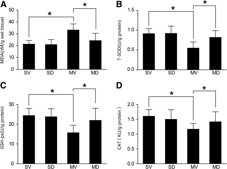
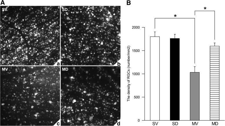
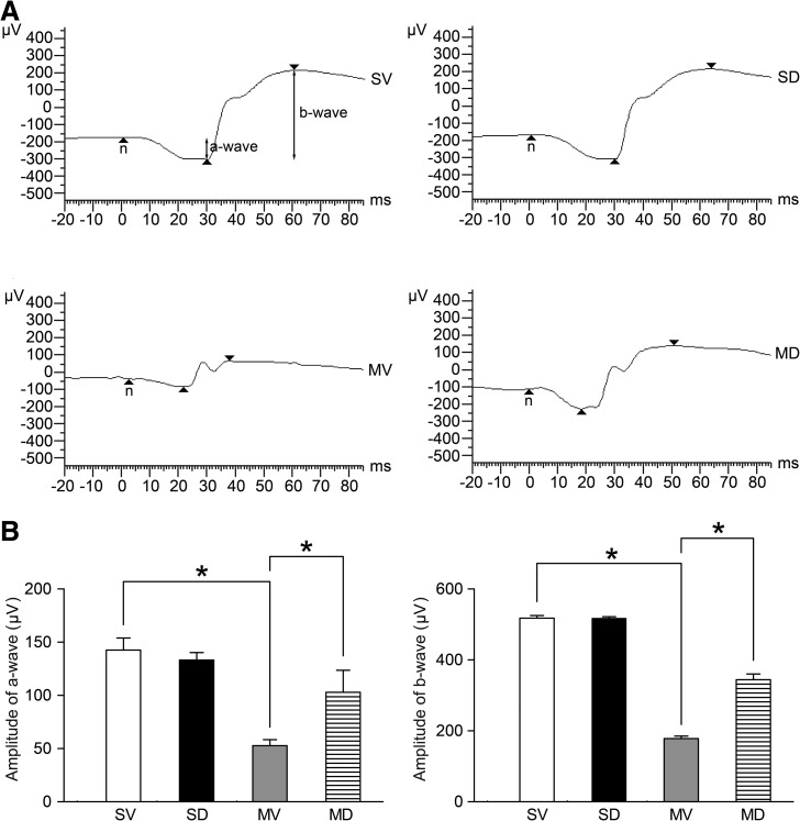
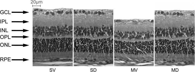
Methods: Retinal I/R injury was induced in the rats by elevating the IOP to 110 mmHg for 60 min. Diosmin (100 mg/kg) or vehicle solution was administered intragastrically 30 min before the onset of ischemia and then daily after I/R injury until the animals were sacrificed. The levels of malondialdehyde (MDA) and the activities of total-superoxide dismutase (T-SOD), glutathione peroxidase (GSH-Px), and catalase (CAT) in the retinal tissues were determined 24 h after I/R injury. At 7 days post-I/R injury, electroretinograms (ERGs) were recorded, and the density of surviving retinal ganglion cells (RGCs) was estimated by counting retrograde tracer-labeled cells in whole-mounted retinas. Retinal histological changes were also examined and quantified using light microscopy.
Results: Diosmin significantly decreased the MDA levels and increased the activities of T-SOD, GSH-Px, and CAT in the retina of rats compared with the ischemia group (P<0.05), and suppressed the I/R-induced reduction in the a- and b-wave amplitudes of the ERG (P<0.05). The thickness of the entire retina, inner nuclear layer, inner plexiform layer, and outer retinal layer and the number of cells in the ganglion cell layer were significantly less after I/R injury (P<0.05), and diosmin remarkably ameliorated these changes on retinal morphology. Diosmin also attenuated the I/R-induced loss of RGCs of the rat retina (P<0.05).
Conclusion: Diosmin protected the retina from I/R injury, possibly via a mechanism involving the regulation of oxidative parameters.
Figures
References
-
- Osborne N.N. Casson R.J. Wood J.P. Chidlow G. Graham M. Melena J. Retinal ischemia: mechanisms of damage and potential therapeutic strategies. Prog. Retin Eye Res. 2004;23:91–147. - PubMed
-
- Louzada-Junior P. Dias J.J. Santos W.F. Lachat J.J. Bradford H.F. Coutinho-Netto J. Glutamate release in experimental ischaemia of the retina: an approach using microdialysis. J. Neurochem. 1992;59:358–363. - PubMed
-
- Dreyer E.B. A proposed role for excitotoxicity in glaucoma. J. Glaucoma. 1998;7:62–67. - PubMed
-
- Li S.Y. Fu Z.J. Ma H. Jang W.C. So K.F. Wong D. Lo A.C. Effect of lutein on retinal neurons and oxidative stress in a model of acute retinal ischemia/reperfusion. Invest. Ophthalmol. Vis. Sci. 2009;50:836–843. - PubMed
-
- Zimmerman B.J. Granger D.N. Oxygen free radicals and the gastrointestinal tract: role in ischemia-reperfusion injury. Hepatogastroenterology. 1994;41:337–342. - PubMed
Publication types
MeSH terms
Substances
LinkOut - more resources
Full Text Sources
Medical
Miscellaneous
