

Anti-caveolin-1 antibodies as anti-prostate cancer therapeutics
- PMID: 22509911
- PMCID: PMC3326273
- DOI: 10.1089/hyb.2011.0100
Anti-caveolin-1 antibodies as anti-prostate cancer therapeutics
Abstract
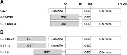
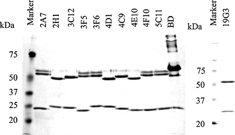
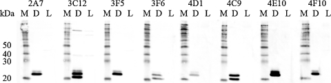
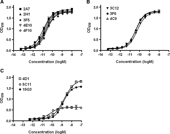
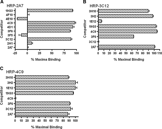
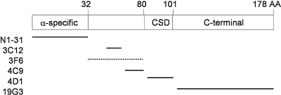
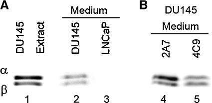
Caveolae are critical cell surface structures important in coordinated cell signaling and endocytosis. One of the major proteins of caveolae is caveolin 1 (Cav-1). Cellular levels of Cav-1 are associated with cancer progression. In prostate cancer cells, levels of Cav-1 are positively correlated with tumor progression and metastasis. Cav-1 can be secreted by prostate cancer cells into the microenvironment and triggers proliferation and anti-apoptosis of the tumor and tumor endothelial cells. Clinical studies have shown increased serum Cav-1 levels in patients with poor prognosis. In tissue culture and animal model experiments, blocking secreted Cav-1 by polyclonal antibodies inhibits tumor cell growth. Cav-1 is therefore a potential therapeutic target for prostate cancer treatment. In this study, we used Cav-1 knock-out mice as hosts to produce monoclonal anti-Cav-1 antibodies. A total of 11 hybridoma cell lines were selected for their ability to produce antibodies that bound GST-Cav-1 but not GST on glutathione-coated ELISA plates. Further screening with ELISAs using GST-Cav-1 fragments on GSH-coated plates classified these antibodies into four groups: N1-31 with five antibodies binds the far N-terminus between amino acids 1 and 31; N32-80 with three antibodies binds between amino acids 32 and 80; CSD with two antibodies potentially bind the scaffolding domain (amino acids 80-101); and Cav-1-C with 1 antibody binds parts of the C-terminal half. Binding affinities (Kd) of these antibodies to soluble Cav-1 ranged from 10(-11) to 10(-8) M. Binding competition experiments revealed that these antibodies recognized a total of six different epitopes on Cav-1. Potency of these antibodies to neutralize Cav-1-mediated signaling pathways in cultured cells and in animal models will be tested. A selected monoclonal antibody will then be humanized and be further developed into a potential anti-prostate cancer therapeutic.
Figures

Similar articles
-
Stromal antiapoptotic paracrine loop in perineural invasion of prostatic carcinoma.Cancer Res. 2006 May 15;66(10):5159-64. doi: 10.1158/0008-5472.CAN-05-1847. Cancer Res. 2006. PMID: 16707439
-
Functional analysis of secreted caveolin-1 in mouse models of prostate cancer progression.Mol Cancer Res. 2009 Sep;7(9):1446-55. doi: 10.1158/1541-7786.MCR-09-0071. Epub 2009 Sep 8. Mol Cancer Res. 2009. PMID: 19737975 Free PMC article.
-
Development of an immunoassay for serum caveolin-1: a novel biomarker for prostate cancer.Clin Cancer Res. 2003 Sep 1;9(10 Pt 1):3653-9. Clin Cancer Res. 2003. PMID: 14506154
-
The role of caveolin-1 in prostate cancer: clinical implications.Prostate Cancer Prostatic Dis. 2010 Mar;13(1):6-11. doi: 10.1038/pcan.2009.29. Epub 2009 Jul 7. Prostate Cancer Prostatic Dis. 2010. PMID: 19581923 Free PMC article. Review.
-
[Caveolin-1: a novel biomarker for prostate cancer].Zhonghua Nan Ke Xue. 2012 Jul;18(7):635-8. Zhonghua Nan Ke Xue. 2012. PMID: 22994052 Review. Chinese.
Cited by
-
Caveolin-1 is a risk factor for postsurgery metastasis in preclinical melanoma models.Melanoma Res. 2014 Apr;24(2):108-19. doi: 10.1097/CMR.0000000000000046. Melanoma Res. 2014. PMID: 24500501 Free PMC article.
-
Caveola-forming proteins caveolin-1 and PTRF in prostate cancer.Nat Rev Urol. 2013 Sep;10(9):529-36. doi: 10.1038/nrurol.2013.168. Epub 2013 Aug 13. Nat Rev Urol. 2013. PMID: 23938946 Review.
-
1,25 Dihydroxyvitamin D3 uptake is localized at caveolae and requires caveolar function.J Biomed Nanotechnol. 2013 Oct;9(10):1707-15. doi: 10.1166/jbn.2013.1662. J Biomed Nanotechnol. 2013. PMID: 24015500 Free PMC article.
-
Endothelial Caveolin-1 regulates the radiation response of epithelial prostate tumors.Oncogenesis. 2015 May 18;4(5):e148. doi: 10.1038/oncsis.2015.9. Oncogenesis. 2015. PMID: 25985209 Free PMC article.
-
Caveolins as Regulators of Stress Adaptation.Mol Pharmacol. 2018 Apr;93(4):277-285. doi: 10.1124/mol.117.111237. Epub 2018 Jan 22. Mol Pharmacol. 2018. PMID: 29358220 Free PMC article. Review.
References
-
- Schlegel A. Volonte D. Engelman JA. Galbiati F. Mehta P. Zhang XL. Scherer PE. Lisanti MP. Crowded little caves: structure and function of caveolae. Cell Signal. 1998;10:457–463. - PubMed
-
- Schlegel A. Pestell RG. Lisanti MP. Caveolins in cholesterol trafficking and signal transduction: implications for human disease. Front Biosci. 2000;5:D929–937. - PubMed
-
- Okamoto T. Schlegel A. Scherer PE. Lisanti MP. Caveolins, a family of scaffolding proteins for organizing “preassembled signaling complexes” at the plasma membrane. J Biol Chem. 1998;273:5419–5422. - PubMed
-
- Razani B. Schlegel A. Liu J. Lisanti MP. Caveolin-1, a putative tumour suppressor gene. Biochem Soc Trans. 2001;29:494–499. - PubMed
Publication types
MeSH terms
Substances
Grants and funding
LinkOut - more resources
Full Text Sources
Other Literature Sources
Medical
Molecular Biology Databases
Research Materials
