. 2012 Aug;143(2):469-80.
doi: 10.1053/j.gastro.2012.04.011. Epub 2012 Apr 14.

Induced Mist1 expression promotes remodeling of mouse pancreatic acinar cells

Affiliations

Induced Mist1 expression promotes remodeling of mouse pancreatic acinar cells

Daniel Direnzo et al. Gastroenterology. 2012 Aug.

Abstract

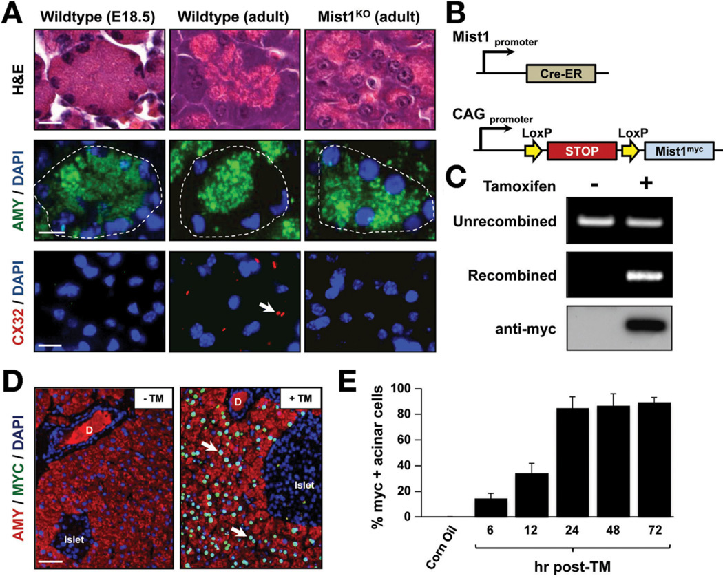
Background & aims: Early embryogenesis involves cell fate decisions that define the body axes and establish pools of progenitor cells. Development does not stop once lineages are specified; cells continue to undergo specific maturation events, and changes in gene expression patterns lead to their unique physiological functions. Secretory pancreatic acinar cells mature postnatally to synthesize large amounts of protein, polarize, and communicate with other cells. The transcription factor MIST1 is expressed by only secretory cells and regulates maturation events. MIST1-deficient acinar cells in mice do not establish apical-basal polarity, properly position zymogen granules, or communicate with adjacent cells, disrupting pancreatic function. We investigated whether MIST1 directly induces and maintains the mature phenotype of acinar cells.

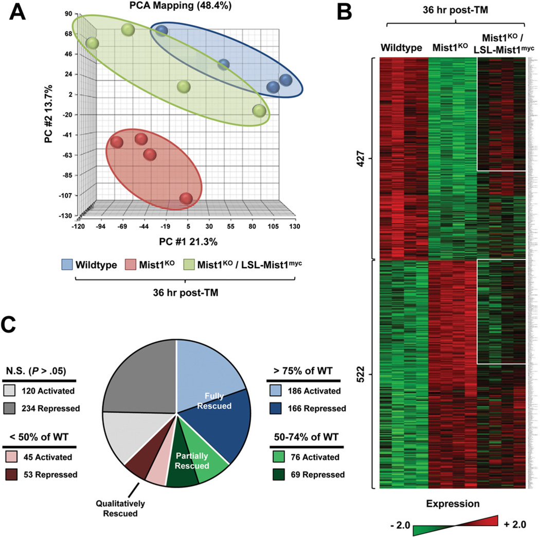
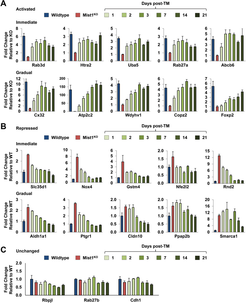
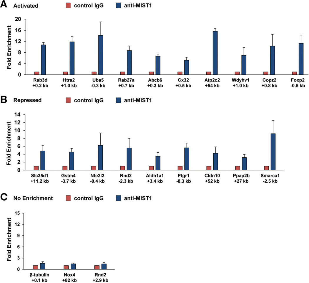
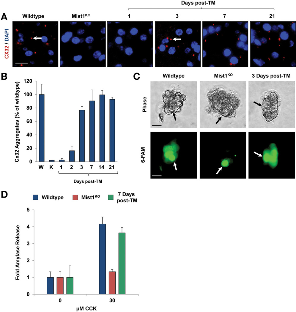
Methods: We analyzed the effects of Cre-mediated expression of Mist1 in adult Mist1-deficient (Mist1(KO)) mice. Pancreatic tissues were collected and analyzed by light and electron microscopy, immunohistochemistry, real-time polymerase chain reaction analysis, and chromatin immunoprecipitation. Primary acini were isolated from mice and analyzed in amylase secretion assays.

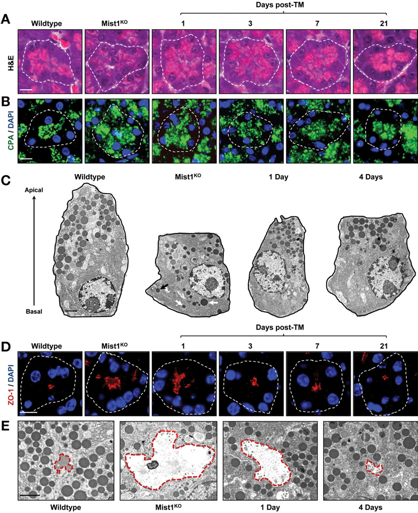
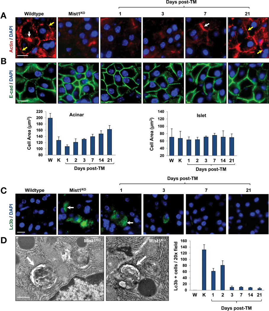
Results: Induced expression of Mist1 in adult Mist1(KO) mice restored wild-type gene expression patterns in acinar cells. The acinar cells changed phenotypes, establishing apical-basal polarity, increasing the size of zymogen granules, reorganizing the cytoskeletal network, communicating intercellularly (by synthesizing gap junctions), and undergoing exocytosis.

Conclusions: The exocrine pancreas of adult mice can be remodeled by re-expression of the transcription factor MIST1. MIST1 regulates acinar cell maturation and might be used to repair damaged pancreata in patients with pancreatic disorders.


Conflict of interest statement

Conflicts of interest

The authors disclose no conflicts.

Figures

Figure 1
Figure 1
Characterization of the LSL-Mist1myc transgenic line. (A) Standard H&E, amylase (AMY), and Cx32 immunofluorescence depicts adult Mist1KO acini showing similarities to embryonic cells, in contrast to adult, fully developed WT acinar cells. Arrows, Cx32 gap junctions. E, embryonic day. (B) Schematic of the Mist1Cre-ER genomic locus and LSL-Mist1myc transgene. (C) Genomic PCR and immunoblots reveal the TM-restricted recombination and expression of the LSL-Mist1myc transgene. (D) Upon TM, only acinar cells express nuclear MIST1myc.protein (arrows). D, duct. (E) Time-course of MIST1myc expression after TM treatment. Scale bars: (A) 10 µm; (D) 50 µm.
Figure 2
Figure 2
Expression of LSL-Mist1myc for 36 hours induces extensive changes in gene expression. (A) Principle component analysis (PCA) of WT, Mist1KO, and Mist1KO/LSLMist1myc mice after TM. Each small circle represents an individual animal. See the Supplementary Materials and Methods section for details. (B) Representative heat map of 4 individual WT, Mist1KO, and Mist1KO/LSL-Mist1myc pancreas RNA samples. The 949 genes that showed a significant difference in expression (P < .05; fold-change, >1.5) between WT and Mist1KO are indicated. White boxes represent fully and partially rescued genes. (C) Pie chart illustrating the degree of recovery for the 949 identified genes in Mist1KO/LSL-Mist1myc mice after TM. See the text for details.
Figure 3
Figure 3
Long-term expression of LSL-Mist1myc further influences gene expression patterns in pancreatic acinar cells. (A and B) Mist1KO/LSL-Mist1myc mice (3 mice per time point) were given a single dose of TM and then RNA was harvested at the indicated times. Reverse-transcription quantitative PCR was used to compare the expression profiles of genes from WT, Mist1KO, and Mist1KO/LSL-Mist1myc pancreata. (C) Control genes Rbpjl, Rab27b, and Cdh1 show no significant changes in gene expression over this time-course.
Figure 4
Figure 4
Chromatin immunoprecipitation assays confirm MIST1 interaction with target genes. (A and B) Pancreas chromatin from 3 WT mice was processed for ChIP assays using anti-IgG (control) or anti-MIST1. In most cases, MIST1 binding was enriched significantly in the anti-Mist1 groups. (C) Notably, no enrichment was observed with control genes (β-tubulin) or at alternative sites within target genes (eg, Rnd2 +2.9 kb). In addition, Nox4 showed no MIST1 binding at the predicted MIST1 binding site. The positions of the amplicon regions with respect to each gene are indicated below the graphs.
Figure 5
Figure 5
Induced MIST1myc rescues the Mist1KO phenotype in adult acinar cells. (A and B) H&E and carboxypeptidase (CPA) immunofluorescence reveals the organization of zymogen granules within individual acini units of WT, Mist1KO, and Mist1KO/LSL-Mist1myc pancreata 1, 3, 7, and 21 days after TM. (C) Transmission electron microscopy views of individual acinar cells showing the mislocalized zymogen granules (arrows) in Mist1KO samples. (D and E) Analysis of lumen size in WT, Mist1KO, and Mist1KO/LSL-Mist1myc acini by zonula occludens 1 (ZO-1) immunofluorescence and transmission electron microscopy. White outlines, individual acini; red outlines, individual lumens. DAPI, 4′,6-diamidino-2-phenylindole. Scale bars: (A, B, and D) 10 µm; (C and E) 2 µm.
Figure 6
Figure 6
Induced MIST1myc rescues the actin cytoskeleton and cell size defects of Mist1KO acinar cells. (A) Anti–β-actin immunofluorescence showing restoration of the terminal (white arrows) and cortical (yellow arrows) actin webs after TM-induced MIST1myc expression. (B) E-cadherin immunofluorescence and quantification of acinar and islet cell areas over the TM time course. (C and D) LC3B immunofluorescence and TEM reveal that Mist1KO acinar cells show high levels of autophagic vacuoles, whereas induced MIST1myc expression reduces their presence toWT levels. (C) LC3B-positive cells (arrows); (D) individual autophagic vesicles (arrows). Scale bars: (A, B, and C) 10 µm; (D) 0.5 µm. DAPI, 4′,6-diamidino-2-phenylindole; E-cad, E-cadherin.
Figure 7
Figure 7
Cx32-containing gap junctions, intercellular communication, and regulated exocytosis are rescued by MIST1myc expression. (A and B) Cx32-containing gap junctions are produced upon Mist1myc expression. Arrows show gap junction complexes. (C) Single-cell injections of 6-carboxyfluorescein (6-FAM) into isolated acini reveal that intercellular transfer of 6-FAM into neighboring acinar cells occurs after MIST1myc induction. Arrows show the injected cell. (D) Quantification of amylase secretion from WT, Mist1KO, and Mist1KO/LSL-Mist1myc acini after cholecystokinin (CCK) stimulation. The defect in secretion observed with Mist1KO acinar cells is reversed completely by induced LSL-Mist1myc expression. Scale bars: (A) 10 µm; (C) 20 µm. DAPI, 4′,6-diamidino-2-phenylindole.

References

    1. de Assis GF, Cestari TM, Sesso A, et al. Post-natal maturation of acinar cells of the guinea pig pancreas: an ultrastructural morphometric study. Anat Histol Embryol. 2003;32:36–41. - PubMed
    1. Jamieson JD, Gorelick FS, Chang A. Development of secretagogue responsiveness in the pancreas. Scand J Gastroenterol Suppl. 1988;151:98–103. - PubMed
    1. Rovira M, Delaspre F, Massumi M, et al. Murine embryonic stem cell-derived pancreatic acinar cells recapitulate features of early pancreatic differentiation. Gastroenterology. 2008;135:1301–1310. 1310, e1–e5. - PMC - PubMed
    1. Johnson CL, Kowalik AS, Rajakumar N, et al. Mist1 is necessary for the establishment of granule organization in serous exocrine cells of the gastrointestinal tract. Mech Dev. 2004;121:261–272. - PubMed
    1. Pin CL, Rukstalis JM, Johnson C, et al. The bHLH transcription factor Mist1 is required to maintain exocrine pancreas cell organization and acinar cell identity. J Cell Biol. 2001;155:519–530. - PMC - PubMed

Publication types

MeSH terms

Substances