. 2012 May 1;11(9):1818-26.
doi: 10.4161/cc.20208. Epub 2012 May 1.

Decreased translation of p21waf1 mRNA causes attenuated p53 signaling in some p53 wild-type tumors

Affiliations

Decreased translation of p21waf1 mRNA causes attenuated p53 signaling in some p53 wild-type tumors

Li-Ju Chang et al. Cell Cycle. .

Abstract

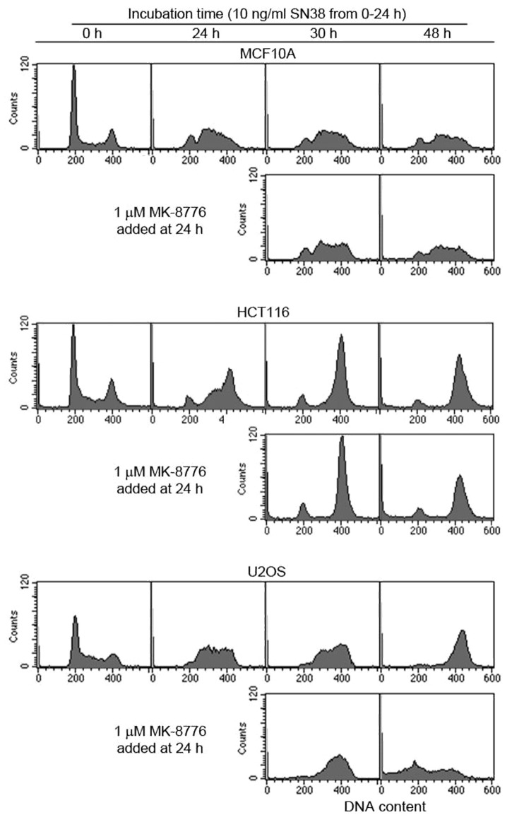
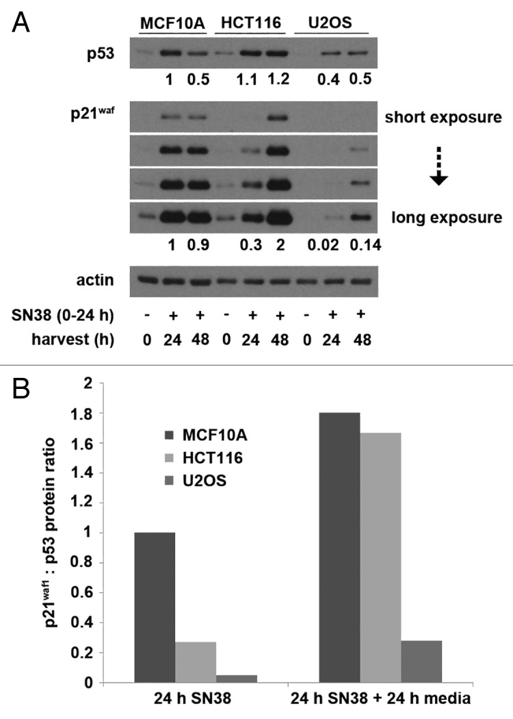
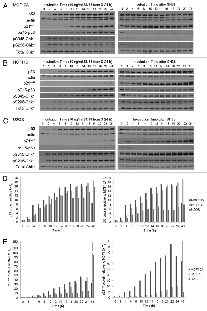
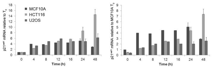
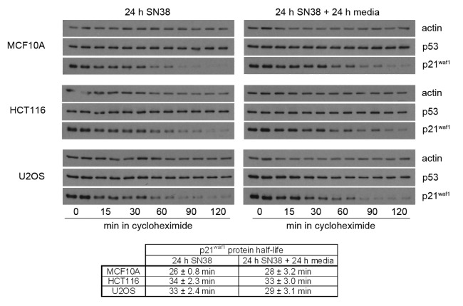
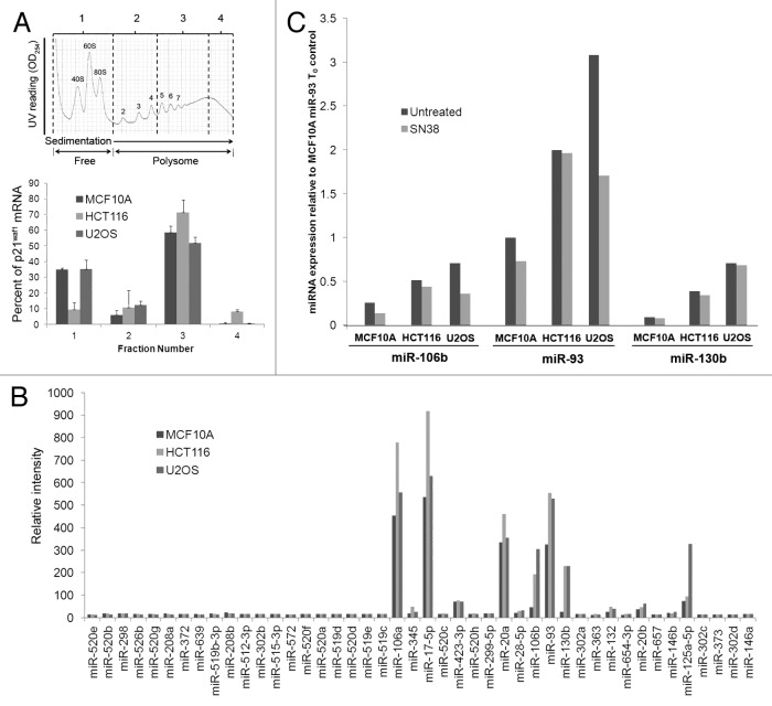
DNA damage induces cell cycle arrest through both Chk1 and the p53 tumor suppressor protein, the latter arresting cells through induction of p21(waf1) protein. Arrest permits cells to repair the damage and recover. The frequent loss of p53 in tumor cells makes them more dependent on Chk1 for arrest and survival. However, some p53 wild type tumor cell lines, such as HCT116 and U2OS, are also sensitive to inhibition of Chk1 due to attenuated p21(waf1) induction upon DNA damage. The purpose of this study is to determine the cause of this attenuated p21(waf1) protein induction. We find that neither the induction of p21(waf1) mRNA nor protein half-life is sufficient to explain the low p21(waf1) protein levels in HCT116 and U2OS cells. The induced mRNA associates with polysomes but little protein is made suggesting these two cell lines have a reduced rate of p21(waf1) mRNA translation. This represents a novel mechanism for disruption of the p53-p21(waf1) pathway as currently known mechanisms involve either mutation of p53 or reduction of p53 protein levels. As a consequence, this attenuated p21(waf1) expression may render some p53 wild type tumors sensitive to a combination of DNA damage plus checkpoint inhibition.


Figures

None
Figure 1. Comparison of the efficacy of MK-8776 to abrogate SN38-induced S and G2 arrest in p53 wild-type cell lines. Cell were incubated with 10 ng/ml SN38 for 24 h and then incubated in either media with or without 1 µM MK-8776. Cells were harvested and assayed for DNA content by flow cytometry.
None
Figure 2. SN38-induced p53 and p21waf1 protein levels. (A) Cells were incubated with 10 ng/ml SN38 for 24 h and then released from SN38 for 24 h. The levels of p53 and p21waf1 protein were assessed by western blotting. Numerical values compare expression level to the 24 h-treated MCF10A cells. (B) The ratio of p21waf1 to p53 protein levels after 24 h of SN38 and after an additional 24 h in fresh medium were compared with that of MCF10A cells after 24 h of SN38.
None
Figure 3. Kinetics of p53 and p21waf1 protein expression following SN38 treatment. (A) MCF10A, (B) HCT116 and (C) U2OS cells were incubated with 10 ng/ml SN38 from 0–24 h. The drug was removed and cells incubated for an additional 24 h in fresh medium. Cells were harvested at the indicated times and proteins detected by western blotting. (D) p53 and (e) p21waf1 protein levels were quantified by densitometry of multiple exposures of western blots and from comparison to a standard curve generated for each antigen. The left panels show protein induction compared with the untreated control of each cell line. The right panels show protein induction compared with the level in untreated MCF10A cells.
None
Figure 4. Kinetics of p21waf1 mRNA induction by SN38 treatment. Cells were incubated with 10 ng/ml SN38 from 0–24 h. The drug was removed and cells were incubated for an additional 24 h in fresh medium. Cells were harvested at the indicated times and p21waf1 mRNA quantified by RT-qPCR. GAPDH was used as an internal control. The left panel shows mRNA induction compared with the untreated control of each cell line. The right panel shows mRNA induction compared with the untreated control of MCF10A. The error bars represent SE of at least three independent experiments.
None
Figure 5. p21waf1 protein half-life following SN38 treatment. Cells were incubated with 10 ng/ml SN38 for 24 h or for an additional 24 h in fresh medium. Cells were then incubated with the protein synthesis inhibitor cyclohexmide (10 µg/ml) for the indicated times. The half-lives are presented as the means ± SE for three independent experiments.
None
Figure 6. Inhibition of p21waf1 mRNA translation. (A) Cells were incubated with 10 ng/ml SN38 for 24 h and then collected for polysome profiling. Samples were divided into four fractions. Fraction one includes the free mRNAs and monosomes, while fractions two, three and four include polysome of increasing size. The bars represent the range of two independent experiments. (B) Cells were incubated with 10 ng/ml SN38 for 24 h and analyzed for levels of 44 miRNAs shown to affect p21waf1 protein levels. (C) Cells were left untreated or incubated with 10 ng/ml SN38 for 24 h. Changes in miR-106b, miR-93 and miR-130b levels were assessed by real-time PCR.

References

    1. Melchionna R, Chen XB, Blasina A, McGowan CH. Threonine 68 is required for radiation-induced phosphorylation and activation of Cds1. Nat Cell Biol. 2000;2:762–5. doi: 10.1038/35036406. - DOI - PubMed
    1. Jazayeri A, Falck J, Lukas C, Bartek J, Smith GC, Lukas J, et al. ATM- and cell cycle-dependent regulation of ATR in response to DNA double-strand breaks. Nat Cell Biol. 2006;8:37–45. doi: 10.1038/ncb1337. - DOI - PubMed
    1. Zhao H, Piwnica-Worms H. ATR-mediated checkpoint pathways regulate phosphorylation and activation of human Chk1. Mol Cell Biol. 2001;21:4129–39. doi: 10.1128/MCB.21.13.4129-4139.2001. - DOI - PMC - PubMed
    1. Clarke CA, Clarke PR. DNA-dependent phosphorylation of Chk1 and Claspin in a human cell-free system. Biochem J. 2005;388:705–12. doi: 10.1042/BJ20041966. - DOI - PMC - PubMed
    1. Niida H, Nakanishi M. DNA damage checkpoints in mammals. Mutagenesis. 2006;21:3–9. doi: 10.1093/mutage/gei063. - DOI - PubMed

Publication types

MeSH terms