

Anesthesia and the quantitative evaluation of neurovascular coupling
- PMID: 22510601
- PMCID: PMC3390804
- DOI: 10.1038/jcbfm.2012.50
Anesthesia and the quantitative evaluation of neurovascular coupling
Abstract
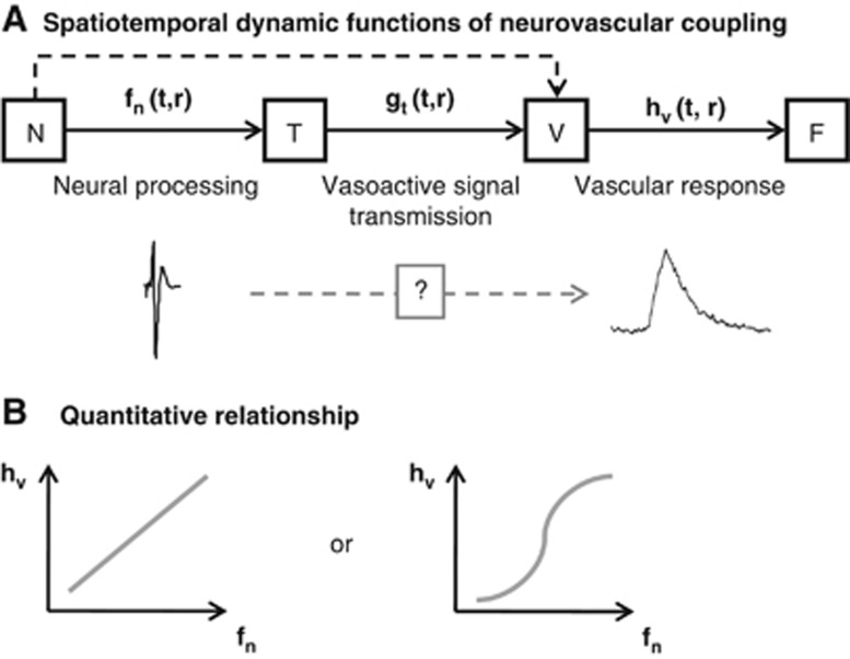
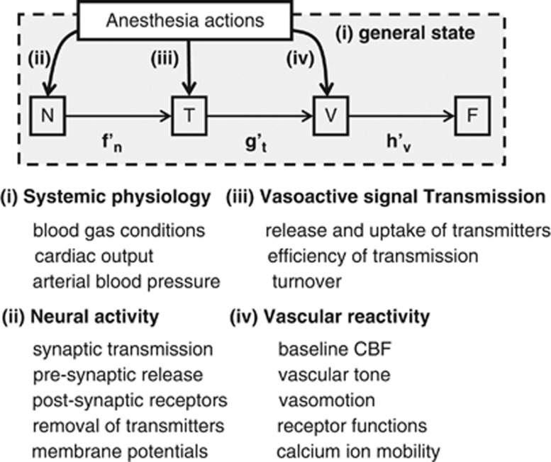
Anesthesia has broad actions that include changing neuronal excitability, vascular reactivity, and other baseline physiologies and eventually modifies the neurovascular coupling relationship. Here, we review the effects of anesthesia on the spatial propagation, temporal dynamics, and quantitative relationship between the neural and vascular responses to cortical stimulation. Previous studies have shown that the onset latency of evoked cerebral blood flow (CBF) changes is relatively consistent across anesthesia conditions compared with variations in the time-to-peak. This finding indicates that the mechanism of vasodilation onset is less dependent on anesthesia interference, while vasodilation dynamics are subject to this interference. The quantitative coupling relationship is largely influenced by the type and dosage of anesthesia, including the actions on neural processing, vasoactive signal transmission, and vascular reactivity. The effects of anesthesia on the spatial gap between the neural and vascular response regions are not fully understood and require further attention to elucidate the mechanism of vascular control of CBF supply to the underlying focal and surrounding neural activity. The in-depth understanding of the anesthesia actions on neurovascular elements allows for better decision-making regarding the anesthetics used in specific models for neurovascular experiments and may also help elucidate the signal source issues in hemodynamic-based neuroimaging techniques.
Figures
References
-
- Akgören N, Dalgaard P, Lauritzen M. Cerebral blood flow increases evoked by electrical stimulation of rat cerebellar cortex: relation to excitatory synaptic activity and nitric oxide synthesis. Brain Res. 1996;710:204–214. - PubMed
-
- Altura BM, Altura BT, Carella A, Turlapaty PD, Weinberg J. Vascular smooth muscle and general anesthetics. Fed Proc. 1980;39:1584–1591. - PubMed
-
- Ances BM, Zarahn E, Greenberg JH, Detre JA. Coupling of neural activation to blood flow in the somatosensory cortex of rats is time-intensity separable, but not linear. J Cereb Blood Flow Metab. 2000;20:921–930. - PubMed
-
- Arfors KE, Arturson G, Malmberg P. Effect of prolonged chloralose anesthesia on acid-base balance and cardiovascular functions in dogs. Acta Physiol Scand. 1971;81:47–53. - PubMed
Publication types
MeSH terms
Substances
LinkOut - more resources
Full Text Sources
Medical
Miscellaneous
