. 2012 Apr 17:3:788.
doi: 10.1038/ncomms1773.

Combinatorial targeting and discovery of ligand-receptors in organelles of mammalian cells

Affiliations
Free PMC article

Combinatorial targeting and discovery of ligand-receptors in organelles of mammalian cells

Roberto Rangel et al. Nat Commun. .
Free PMC article

Abstract

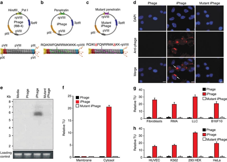
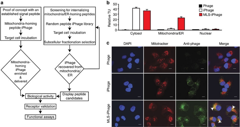
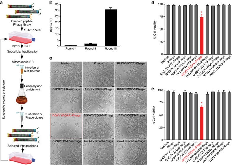
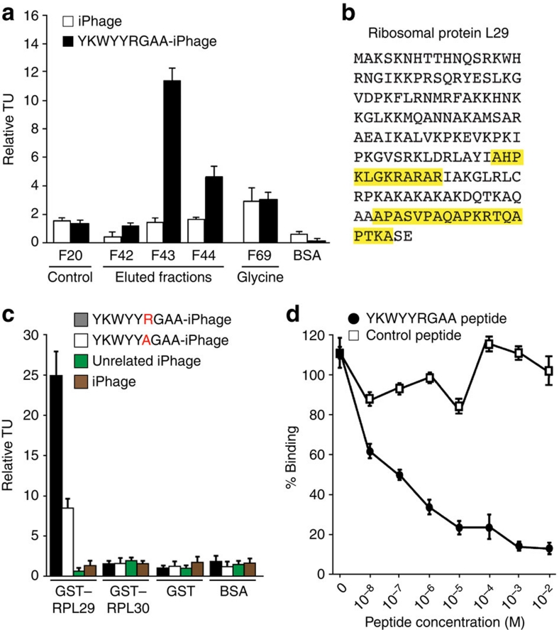
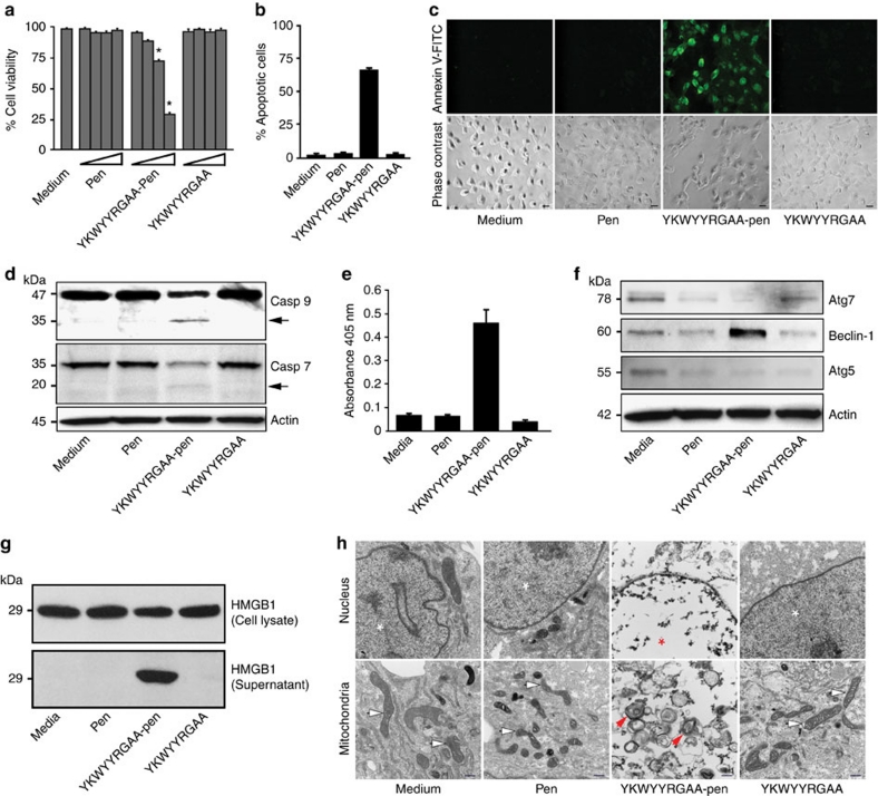
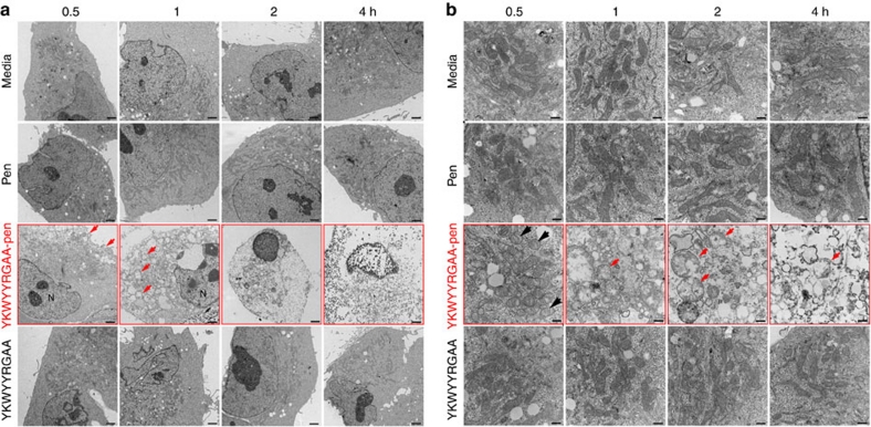
Phage display screening allows the study of functional protein-protein interactions at the cell surface, but investigating intracellular organelles remains a challenge. Here we introduce internalizing-phage libraries to identify clones that enter mammalian cells through a receptor-independent mechanism and target-specific organelles as a tool to select ligand peptides and identify their intracellular receptors. We demonstrate that penetratin, an antennapedia-derived peptide, can be displayed on the phage envelope and mediate receptor-independent uptake of internalizing phage into cells. We also show that an internalizing-phage construct displaying an established mitochondria-specific localization signal targets mitochondria, and that an internalizing-phage random peptide library selects for peptide motifs that localize to different intracellular compartments. As a proof-of-concept, we demonstrate that one such peptide, if chemically fused to penetratin, is internalized receptor-independently, localizes to mitochondria, and promotes cell death. This combinatorial platform technology has potential applications in cell biology and drug development.


Conflict of interest statement

The University of Texas and some of its researchers (W.A. and R.P.) have equity in Alvos Therapeutics and in Ablaris Therapeutics, which are subjected to certain restrictions under university policy; the university manages and monitors the terms of these arrangements in accordance with its conflict-of-interest policy.

Figures

Figure 1
Figure 1. Display of pen on rpVIII mediates receptor-independent cell internalization.
(a) Upper: parental f88-4 phage vector contains two capsid genes encoding a wild-type (wt) protein VIII (pVIII, depicted in grey) and a recombinant protein VIII (rpVIII; depicted in green). The recombinant gene VIII contains a foreign DNA insert with a HindIII and a PstI cloning site (depicted in red). TetR, tetracycline resistance gene. Lower: representation of the assembled phage particle expressing only the wt major coat protein pVIII (grey); pIII, minor coat protein (orange); pVI protein (blue); pVII protein (red), and pIX protein (yellow). (b) Upper: annealed oligonucleotides encoding the pen peptide were cloned in frame with the recombinant gene VIII. Lower: iPhage particles displaying the pen peptide motif (RQIKIWFQNRRMKWKK) at the amino terminus of the rpVIII (green). (c) Upper: the mutant iPhage genome has nucleotide substitutions in the pen sequence replacing tryptophan (W) by alanine (A) residues (underlined in red). Lower: representation of the assembled mutant iPhage displaying mutant pen on rpVIII protein (purple). (d) Immunofluorescence of KS1767 cells shows internalized viral particles only in cells incubated with iPhage. The nuclear stain DAPI emits blue fluorescence, and internalized phage particles were detected with conjugated antibodies (red fluorescence). Scale bar, 100 μm. (e) Phage genomic DNA was detected only in KS1767 cells incubated with iPhage particles (Southern blot, upper panel). Total genomic DNA stained with ethidium bromide served as a loading control (lower panel). (f) iPhage particles are internalized and viable in the cytosol and undetected in the membrane fraction of KS1767 cells. Bars represent mean values for TU recovered from the cytosol±s.e.m., from triplicates. Phage internalization by various types of cultured cells (g) mouse and (h) human. RMA (Rauscher murine leukaemia virus antigen) lymphoma, LLC (Lewis lung carcinoma), B16F10 (Melanoma), HUVEC (human umbilical vein endothelial cell), leukaemia (K562), 293HEK (human embryonic kidney) and HeLa cell lines. Internalization assays were run in triplicate; bars represent mean values for phage TU recovered from the cytosol-enriched fraction±s.e.m. from triplicates.
Figure 2
Figure 2. Systematic approach for mammalian organelle targeting.
(a) Organelle targeting and random peptide iPhage library selection flow diagram. (b) Phage displaying the mitochondrial localization signal of cytochrome c oxidase (MLS-iPhage) in the pIII protein is enriched in the mitochondria/ER fraction. Cytosol and nuclear fractions served as internalizing and negative controls, respectively. Bars represent mean values for TU recovered from the mitochondria/ER fraction±s.e.m., from triplicates. (c) Confocal fluorescence microscopy analysis of KS1767 cells exposed to parental phage, iPhage and MLS-iPhage. Intracellular localization was revealed by anti-rabbit Alexa Fluor 488, orange-Mitotracker, and DAPI counterstaining. Scale Bar, 10 μm. Arrows indicate signal co-localization.
Figure 3
Figure 3. Synchronous selection of a random peptide iPhage library is enriched in the mitochondria/ER fraction.
(a) Systematic approach of selection in live KS1767 cells. (b) Phage enrichment after three rounds of selection. Bars represent mean values for phage TU recovered from the mitochondria/ER enriched fraction±s.e.m., from triplicates. (c) Bright-field microscopy of KS1767 cells infected with different peptide-iPhage clones selected from the mitochondria/ER fraction. Scale bar, 100 μm. Cell viability was measured using two independent methods (d) WST-1 and (e) MTT assays. Bars represent mean values of cell viability±s.e.m., from triplicates. The symbol * indicates significant reduction of cell viability (P<0.02). Data were statistically analysed by Student's t-test.
Figure 4
Figure 4. RPL29 is the receptor for the internalizing YKWYYRGAA peptide.
(a) Phage-binding assay on fractions from YKWYYRGAA affinity chromatography. Fractions were immobilized in a 96-well plate. Insertless iPhage and BSA were used as negative controls. Phage binding assays were run in triplicate; bars represent mean values for phage TU recovered from immobilized proteins±s.e.m., from triplicates. (b) RPL29 was identified by mass spectrometry. Amino acids in yellow represent the peptides detected by mass spectrometry. (c) Phage binding to immobilized GST–RPL29 and GST–RPL30 fusion proteins. BSA or GST alone was used as negative controls. Bars represent mean values for phage TU recovered from immobilized proteins±s.e.m., from triplicates. (d) Binding of the YKWYYRGAA-phage to RPL29 was inhibited with the corresponding synthetic peptide. Bars represent mean±s.e.m., from triplicates.
Figure 5
Figure 5. The internalizing YKWYYRGAA peptide activates cell death via ribosomal protein L29.
(a) Cell viability is reduced on exposure of YKWYYRGAA-pen, relative to complete media, pen, and YKWYYRGAA peptide (1, 3, 10 and 30 μM). The symbol * indicates significant reduction of cell viability (P<0.001; Student's t-test). The MTT assay was performed in triplicate. Bars represent mean±s.e.m. (b) Internalization of YKWYYRGAA peptide induces mammalian cell death. Fluorescence-activated cell sorting of annexin V-FITC-positive cells (apoptotic cells). KS1767 cells were treated with peptides at 30 μM for 6 h at 37 °C. Bars represent mean±s.e.m., from triplicates. (c) Annexin V-FITC binds to translocated phophatidylserine on the plasma membrane of cells treated with YKWYYRGAA-pen. Control, pen, and YKWYYRGAA-treated cells did not translocate the apoptotic marker. Scale bar, 100 μm. (d) The internalizing YKWYYRGAA peptide activates the caspases in the human cell line KS1767. Immunoblotting shows cleavage of caspase-7 (Casp 7) and -9 (Casp 9). Actin served as a loading control. Arrows indicate the caspase fragments. (e) Histone-associated DNA fragments are detected only in cells treated with YKWYYRGAA-pen peptide. DNA fragmentation was detected with the Cell death detection ELISA plus kit (Roche). Bars represent mean±s.e.m., from triplicates. (f) Beclin-1 is upregulated after YKWYYRGAA-pen peptide treatment. Beclin 1, autophagy related protein (atg) 5, and atg-7 expression were detected by western blot. (g) Actin served as a loading control YKWYYRGAA-pen induced the translocation of the high-mobility group protein B1 (HMGB1) into the extracellular environment. KS1767 cells were treated with different peptides at 30 μM or with media as indicated. (h) Normal mitochondria change to vesicular form, and the matrix swells (red arrows). Chromatin condensation and fragmentation (red asterisk) are observed only in KS1767 cells exposed to YKWYYRGAA-pen. Normal mitochondria (white arrows) and nuclei (white asterisks) are observed in control-untreated, pen-treated, and YKWYYRGAA-treated cells. Scale bar, 500 nm.
Figure 6
Figure 6. The YKWYYRGAA-pen peptide induces ultrastructural alterations leading to cell death.
(a) Electron microscopy analysis of KS1767 cells untreated (media) and treated with peptides (30 μM) at different time points (0.5, 1, 2, and 4 h). The cells treated only with the YKWYYRGAA-pen peptide suffered ultrastructural alterations as early as 30 min; the plasma membrane and the cytoplasm were extensively dismantled (red arrows) compared with cells treated with peptide controls or media. Scale bar, 2 μm. (b) Intracellular compartments analysis by transmission electron microscopy shows normal-vesicular (black arrow) to swollen mitochondria (red arrow), such structural alterations were only observed in KS1767 cells treated with YKWYYRGAA-pen peptide (30 μM). Normal mitochondria were observed in KS1767 cells treated with control peptides (30 μM) and media. Scale bar, 500 nm.

References

    1. Pasqualini R. & Ruoslahti E. Organ targeting in vivo using phage display peptide libraries. Nature 380, 364–366 (1996). - PubMed
    1. Arap W., Pasqualini R. & Ruoslahti E. Cancer treatment by targeted drug delivery to tumor vasculature in a mouse model. Science 279, 377–380 (1998). - PubMed
    1. Arap W. et al. Steps towards mapping the human vasculature by phage display. Nat. Med. 8, 121–127 (2002). - PubMed
    1. Staquicini F. I., Pasqualini R. & Arap W. Ligand-directed profiling: applications to cancer drug discovery. Expert Opin. Drug Discov. 4, 51–59 (2009). - PubMed
    1. Staquicini F. I. et al. Vascular ligand-receptor mapping by direct combinatorial selection in cancer patients. Proc. Natl Acad. Sci. USA 108, 18637–18642 (2011). - PMC - PubMed

Publication types

MeSH terms