Review
. 2012 May;139(10):1701-11.
doi: 10.1242/dev.068338.

Mechanisms of tissue fusion during development

Affiliations
Review

Mechanisms of tissue fusion during development

Heather J Ray et al. Development. 2012 May.

Abstract

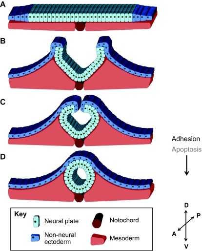
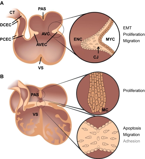
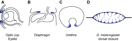
Tissue fusion events during embryonic development are crucial for the correct formation and function of many organs and tissues, including the heart, neural tube, eyes, face and body wall. During tissue fusion, two opposing tissue components approach one another and integrate to form a continuous tissue; disruption of this process leads to a variety of human birth defects. Genetic studies, together with recent advances in the ability to culture developing tissues, have greatly enriched our knowledge of the mechanisms involved in tissue fusion. This review aims to bring together what is currently known about tissue fusion in several developing mammalian organs and highlights some of the questions that remain to be addressed.


Figures

Fig. 1.
Fig. 1.
Tissue fusion during palate development. (A) Fusion during murine primary palate development. At embryonic day 9.5 (E9.5), the frontonasal prominence (yellow) is beginning to develop into the medial nasal prominence (MNP) and the lateral nasal prominence (LNP). Also evident is the maxillary prominence (MxP; green) and the mandibular prominence (MAND; purple). Around embryonic day 10.5 (E10.5), initial tissue fusion occurs between the MNP and the MxP (area between arrows) followed by fusion of the MNP and LNP. (B) Fusion during murine secondary palate development. At E13.5, the palatal shelves (P) are oriented vertically along the tongue (T). By E14, movement of the palatal shelves has brought them into a horizontal position above the tongue. From E14.5 to E16, opposing palatal shelves fuse (area between arrows) to generate the secondary palate. The mechanisms that are known to be necessary (black) or implicated (gray) in primary and secondary palatal fusion are indicated.
Fig. 2.
Fig. 2.
Development and fusion of the neural tube. (A) Thickening of the dorsal neuroepithelium results in formation of the neural plate. (B) The neuroepithelium bends dorsally to form the neural folds. (C) Further bending brings the neural folds in close opposition to each other. Non-neural ectoderm cells cover the edge of the neuroepithelium. (D) Separation of neural and non-neural ectoderm and fusion of these tissues results in formation of the closed neural tube covered by a sheet of ectoderm. Necessary (black lettering) and observed but not required (gray lettering) mechanisms of neural tube fusion are indicated. D, dorsal; V, ventral; A, anterior; P, posterior.
Fig. 3.
Fig. 3.
Fusion during development of the heart. (A) Between E9.5 and E10.5, endocardial cushions (ECs) begin to form across the conotruncus (CT) and the atrioventricular canal (AVC): the proximal conotruncal endocardial cushions (PCECs), the distal conotruncal endocardial cushions (DCECs) and the atrioventricular endocardial cushions (AVECs). The inset provides a closer view of AVEC formation. Endocardial cells (ENC) proliferate, undergo an epithelial-to-mesenchymal transition and migrate into the cardiac jelly (CJ), which is produced by myocardial cells (MYC). (B) Between E12.5 and E13.5, the ECs are in close opposition and begin to fuse. The myocardium-derived primary atrial septum (PAS) and ventricular septum (VS) grow inwards towards the AVEC. The top inset illustrates the mesenchymal cap (MC) that is present at the growing end of both the PAS and the VS. The bottom inset illustrates the fusing ECs as the two cushions come into contact: endothelial integrity at the midline breaks down and the mesenchymal cells migrate across the midline to form ‘mesenchymal bridges’. Necessary (black lettering) and possible (gray lettering) mechanisms involved in EC fusion are indicated.
Fig. 4.
Fig. 4.
Additional tissue fusion events during development. (A) During development of the eye, two tissue fusion events occur: the first drives formation of the optic cup; the second fuses the eyelids together. (B) Tissue fusion occurs to generate the mammalian diaphragm. (C) Opposing epithelia fuse to complete the tubular urethra. (D) In Drosophila melanogaster, fillipodia (blue spikes) extend from opposing epithelia to aid in the dorsal closure fusion event. Tissue fusion events occur between tissues shaded in blue. Adapted, with permission, from Yu et al. (Yu et al., 2010).

References

    1. Abbott B. D. (2010). The etiology of cleft palate: a 50-year search for mechanistic and molecular understanding. Birth Defects Res. B Dev. Reprod. Toxicol. 89, 266–274 - PubMed
    1. Abdul-Aziz N. M., Turmaine M., Greene N. D., Copp A. J. (2009). EphrinA-EphA receptor interactions in mouse spinal neurulation: implications for neural fold fusion. Int. J. Dev. Biol. 53, 559–568 - PubMed
    1. Beverdam A., Brouwer A., Reijnen M., Korving J., Meijlink F. (2001). Severe nasal clefting and abnormal embryonic apoptosis in Alx3/Alx4 double mutant mice. Development 128, 3975–3986 - PubMed
    1. Bhattacherjee V., Horn K. H., Singh S., Webb C. L., Pisano M. M., Greene R. M. (2009). CBP/p300 and associated transcriptional co-activators exhibit distinct expression patterns during murine craniofacial and neural tube development. Int. J. Dev. Biol. 53, 1097–1104 - PMC - PubMed
    1. Blavier L., Lazaryev A., Groffen J., Heisterkamp N., DeClerck Y. A., Kaartinen V. (2001). TGF-beta3-induced palatogenesis requires matrix metalloproteinases. Mol. Biol. Cell 12, 1457–1466 - PMC - PubMed

Publication types