. 2012 May;139(10):1851-62.
doi: 10.1242/dev.075788.

The novel intestinal filament organizer IFO-1 contributes to epithelial integrity in concert with ERM-1 and DLG-1

Affiliations

The novel intestinal filament organizer IFO-1 contributes to epithelial integrity in concert with ERM-1 and DLG-1

Katrin Carberry et al. Development. 2012 May.

Abstract

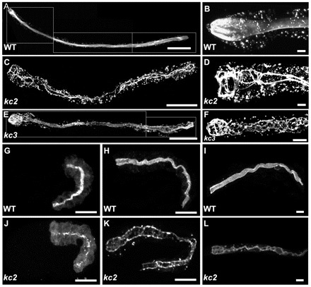
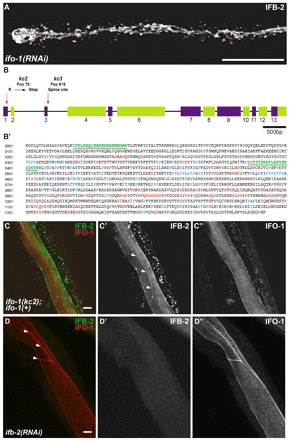
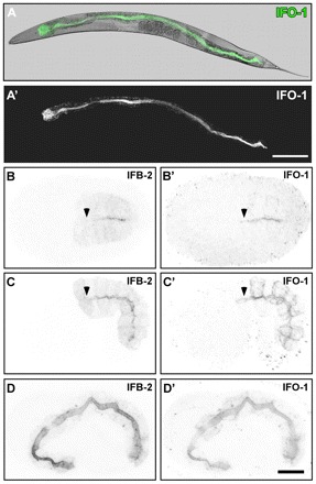
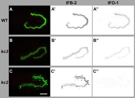
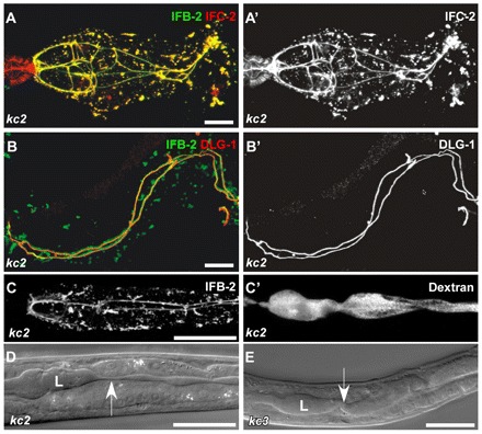
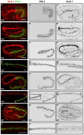
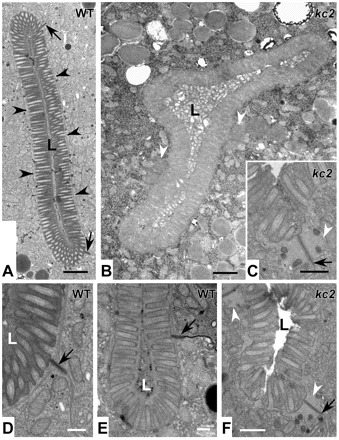
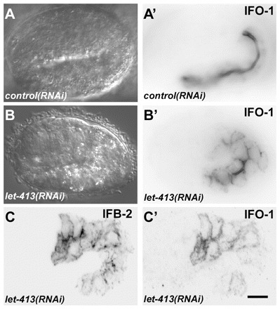
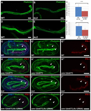
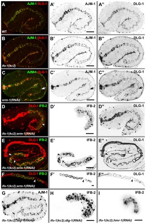
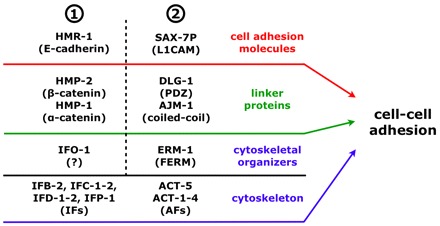
The nematode Caenorhabditis elegans is an excellent model system in which to study in vivo organization and function of the intermediate filament (IF) system for epithelial development and function. Using a transgenic ifb-2::cfp reporter strain, a mutagenesis screen was performed to identify mutants with aberrant expression patterns of the IF protein IFB-2, which is expressed in a dense network at the subapical endotube just below the microvillar brush border of intestinal cells. Two of the isolated alleles (kc2 and kc3) were mapped to the same gene, which we refer to as ifo-1 (intestinal filament organizer). The encoded polypeptide colocalizes with IF proteins and F-actin in the intestine. The apical localization of IFO-1 does not rely on IFB-2 but is dependent on LET-413, a basolateral protein involved in apical junction assembly and maintenance of cell polarity. In mutant worms, IFB-2 and IFC-2 are mislocalized in cytoplasmic granules and accumulate in large aggregates at the C. elegans apical junction (CeAJ) in a DLG-1-dependent fashion. Electron microscopy reveals loss of the prominent endotube and disordered but still intact microvilli. Semiquantitative fluorescence microscopy revealed a significant decrease of F-actin, suggesting a general role of IFO-1 in cytoskeletal organization. Furthermore, downregulation of the cytoskeletal organizer ERM-1 and the adherens junction component DLG-1, each of which leads to F-actin reduction on its own, induces a novel synthetic phenotype in ifo-1 mutants resulting in disruption of the lumen. We conclude that IFO-1 is a multipurpose linker between different cytoskeletal components of the C. elegans intestinal terminal web and contributes to proper epithelial tube formation.


Figures

Fig. 1.
Fig. 1.
EMS mutagenesis induces mislocalization of IFB-2::CFP. (A-L) The fluorescence images show the IFB-2::CFP reporter distribution in WT background (strain BJ52; A,B,G-I) and kc2 (C,D,J-L) and kc3 (E,F) mutant worms of strains BJ133 and BJ134, respectively. (A-F) Adult worms; (G,H,J,K) embryos; and (I,L) L1 larvae. In WT adults, IFB-2::CFP localizes to the subapical perilumenal cytoskeletal network of intestinal cells (A). By contrast, in kc2 mutants IFB-2::CFP forms multiple cytoplasmic aggregates and is almost completely absent from the apical part of the cell except for a characteristic rope ladder-type pattern (C). B and D show magnifications of the anterior parts of WT and kc2 mutant adults, respectively. Young kc3 adults (<3 days) develop a less severe phenotype than kc2 with fewer cytoplasmic aggregates and only some small gaps in the subapical IFB-2::CFP network (E). In older adult worms (>3 days), however, the phenotype becomes more severe, as the gaps become bigger and cytoplasmic aggregates increase in size and number (F). In WT embryos, IFB-2 localizes to the apical membrane domain in mid-morphogenesis (G). A diffuse cytoplasmic staining can be observed which becomes weaker during further development (2-fold stage, H) and is almost undetectable in L1 larvae (I). In kc2 mutants, IFB-2 is never properly localized. During mid-morphogenesis (J) and in a 2.5-fold elongated embryo (K) only a punctate IFB-2 pattern is seen. In the L1 larvae, IFB-2 localizes in a rope ladder pattern at the cell apex and in cytoplasmic aggregates (L). Scale bars: 100 μm in A,C,E; 15 μm in G,H,J,K; 10 μm in B,D,F,I,L.
Fig. 2.
Fig. 2.
kc2 and kc3 are alleles of gene F42C5.10 encoding intestinal filament organizer IFO-1, which colocalizes with IFB-2. (A) Fluorescence image of IFB-2::CFP-expressing reporter strain BJ52 after RNAi against F42C5.10-encoded RNA shows identical phenotype to kc2 mutant (see Fig. 1C,D). (B,B′) Structure of the F42C5.10 ifo-1 gene and encoded polypeptide sequence. The 5789 bp-long gene consists of 13 exons. Mutant allele kc2 carries a C→T mutation at position 79 leading to a stop codon in the first exon, kc3 a G→A mutation at position 919, which is the first position of the 5′ donor splice site of the third intron. The encoded polypeptide, referred to as IFO-1, is 1292 amino acids long. It is rich in histidines (marked in red) and contains a proline-rich region (prolines highlighted in blue). The two peptides (green) used for antibody generation are underlined. (C-C″) Fluorescence microscopy of mutant kc2 worms of strain BJ133 that were microinjected with an ifo-1::yfp rescue construct. The normal-appearing, fully rescued IFB-2::CFP fluorescence in green (false color) co-distributes with the IFO-1::YFP fluorescence (false red color). (D-D″) Fluorescence microscopy of isolated WT intestines after ifb-2(RNAi) that were stained against IFB-2 (false green color) and with purified anti-IFO-1 peptide antibodies (false red color; peptide 2 in B′). Arrows in C′ and D delineate the CeAJ. Scale bars: 50 μm in A; 10 μm in C,D.
Fig. 3.
Fig. 3.
IFO-1 is expressed exclusively in the intestine and localizes to the adlumenal domain together with IFB-2. Fluorescence and DIC pictures of IFO-1::CFP-expressing adult worm (A,A′) and embryos (B-D′) of strain BJ155. (A-A′) IFO-1 fluorescence (green) is detected in adult worms only in the intestine where it localizes apically (overlay of DIC and fluorescence image in A). (B-D′) Embryos co-stained with anti-IFB-2 antibodies show that IFO-1 fluorescence is first detectable during polarization of the intestinal primordium, where it accumulates at the future apical pole together with IFB-2 (B,B′). In mid-morphogenesis, IFO-1 is also present at the lateral cortex that is negative for IFB-2. By contrast, IFB-2 localizes diffusely to the cytoplasm, which is negative for IFO-1 (C,C′). During further development, both proteins continue to accumulate at the apical domain whereas the respective lateral and cytoplasmic fluorescence signals diminish (3-fold embryo in D,D′). Note the absence of anti-IFB-2 signal (arrowheads) in some apical membrane regions that already stain positive for IFO-1::CFP (arrowheads). Scale bars: 100 μm in A′; 10 μm in D’ (for B-D′).
Fig. 4.
Fig. 4.
Peptide antibodies detect IFO-1 exclusively perilumenally in the intestine of WT and at reduced levels of kc3 mutants but not in the intestine of kc2 mutants. (A-C″) The images show immunofluorescence analyses (overlay at left; inverse presentation in middle and right column) of WT (A-A″), kc3 (B-B″) and kc2 (C-C″) 2-fold embryos that were stained against IFB-2 (green) and with purified anti-IFO-1 peptide antibodies (red; peptide 2, see Fig. 2B′). The micrographs that were taken under the same conditions show a decrease of intestinal IFO-1 in kc3 (B″) and a complete loss of the protein in kc2 (C). Scale bar: 10 μm.
Fig. 5.
Fig. 5.
In ifo-1 mutant worms, IF proteins IFB-2 and IFC-2 colocalize and co-distribute with the CeAJ marker DLG-1. The intestinal lumen is perturbed but still tight. (A,A′) Fluorescence of dissected adult ifo-1(kc2) intestines detecting IFB-2::CFP and anti-IFC-2. IFC-2 colocalizes with IFB-2 in cytoplasmic aggregates and at the CeAJ in anterior intestinal cells. Note the presence of IFC-2 but not of IFB-2 in the adjacent pharynx. (B,B′) The re-distributed anti-IFB-2 immunofluorescence colocalizes with anti-DLG-1 immunofluorescence at the CeAJ but shows a more extensive staining adjacent to the junction and in cytoplasmic granules. (C,C′) Fluorescence micrographs depict the disturbed IFB-2::CFP distribution in kc2 mutant worms (strain BJ133) together with TRITC-labeled dextrane that was taken up by feeding. Note that the dextrane is restricted to the altered lumen indicating that the junctional seal is still intact. (D,E) DIC micrographs reveal an abnormal lumen (L) with multiple constrictions (arrow) and dilations in kc2 and kc3 mutant worms (strains BJ133 and BJ134, respectively). Scale bars: 10 μm in A,B; 100 μm in C-E.
Fig. 6.
Fig. 6.
IFB-2 mislocalization in kc2 and kc3 mutants develops differently. Embryos (A-C,E,G) and L1/L2 larvae (D,F,H) were stained with antibodies against IFB-2 (green, A-H; inverted, middle column) and DLG-1 (red, A-H; inverted, right column). (A-C″) In the 1.5-fold stage of kc2 mutants, IFB-2 is localized at the apical part of intestinal cells in a punctate pattern (A-A″), starts to accumulate at the CeAJ at the 2-fold stage but is still present in apical puncta (B-B″) and becomes fully recruited to the CeAJ at the 3-fold stage (C-C″; note increase of cytoplasmic IFB-2 granules in number, especially in the anterior intestine). (D-D″) In kc2 L2, the aggregates further increase and are more evenly distributed throughout the intestine. (E-E″) By contrast, IFB-2 is distributed normally in kc3 embryos (compare with Fig. 3D). (F-F″) In the kc3 L1 stage, small gaps (see magnification in F’) appear in the apical IFB-2-positive network. The gaps increase in size in older worms until the phenotype resembles that of kc2 worms (see Fig. 1). (G-H″) In WT embryos and L1 larvae, the contiguous anti-IFB-2 and anti-DLG-1 signals delineate the endotube and the CeAJ, respectively. Scale bars: 10 μm.
Fig. 7.
Fig. 7.
ifo-1 mutants lack the prominent endotube and present instead large junctional aggregates. Electron micrographs taken from WT N2 (A,D,E) and strain BJ142 ifo-1(kc2) (B,C,F). Note the electron dense endotube in WT (A, black arrowheads), which is absent in kc2 and the presence of large aggregates (white arrowheads in B,C,F) next to the CeAJ (black arrows). In addition, the lumen (L) is distorted in kc2 (B) and the microvilli are less ordered (C,F). They still contain, however, characteristic parallel actin filament bundles. Scale bars: 500 nm in A,B; 250 nm in C-F.
Fig. 8:
Fig. 8:
let-413 RNAi disturbs proper IFO-1 and IFB-2 localization. (A-B′) Embryos of reporter strain BJ186 kcEX28[ifo-1::yfp,myo-3p::mCherry] were treated with dsRNA directed against either mtm-6 (A,A′) or let-413 (B,B′). DIC images (A,B) and corresponding fluorescence micrographs (A′,B′) are shown. Note the typical exclusive apical IFO-1 distribution in the control that contrasts with the circumferential labeling of apical and basolateral cortical domains upon let-413 RNAi treatment. (C,C′) Fluorescence micrographs of 1.75-fold embryo of strain BJ155 kcEx29[ifo-1::cfp, myo-3p::mcherry] depicting antibody staining of anti-IFB-2 in C and IFO-1::CFP fluorescence in C′ upon let-413 RNAi. Note the non-polarized distribution of both. Scale bar: 10 μm.
Fig. 9.
Fig. 9.
ifo-1 mutants show reduced intestinal F-actin and are synthetic with erm-1. (A-F) Detection of F-actin either by phalloidin staining (A,B) or by antibodies against F-actin (D,E) of isolated adult intestines (WT in A,D; ifo-1(kc2) in B,E) reveals a significant reduction in fluorescence (quantification in C,F; P<0.001). Error bars represent s.d. (G-J″) Anti-ERM-1 (blue) and anti-IFB-2 (red) immunofluorescence and phalloidin (green). The position of the intestinal lumen is marked by arrowheads. Representative images were taken under the same conditions. Note normal localization of ERM-1 in kc2 (I) and absence of anti-ERM-1 signal in tm677 (H,J). (G′-J″) Selective reduction of intestinal phalloidin labeling and anti-IFB-2 immunofluorescence (see Results for quantification) in comparison with WT (G′,G″) is detectable in erm-1(tm677) (H′,H″) and in kc2 (I′,I″) embryos. Note the synthetic phenotype resulting in strong reduction of intestinal phalloidin (J’) and IFB-2 (J″) in erm-1(tm677);ifo-1(RNAi) embryos. Scale bars: 10 μm.
Fig. 10.
Fig. 10.
ifo-1 mutants are synthetic with erm-1 and dlg-1 mutants but not with hmr-1 mutants. (A-C″) Anti-AJM-1 (green in A-C; inverted in A′-C′) and anti-DLG-1 (red in A-C; inverted in A″-C″) immunofluorescence in 1.75-fold stages of WT (A-A″), ifo-1(kc2) (B-B″) and after RNAi against erm-1 (C-C″). Arrow indicates constriction of the future lumen. (D-F″) Fluorescence micrographs of IFB-2::CFP (green in D-F; inverted in D′-F′) and anti-DLG-1 signal (red, D-F; inverted D″-F″) in kc2 background of 1.75-fold embryo (D-D″), 3-fold embryo (E-E″) and L1-larva (F-F″) treated with dsRNA against erm-1. (G,H) Immunofluorescence of AJM-1 (G) and IFB-2 (H) in 1.75-fold ifo-1(kc2);dlg-1(RNAi) embryo. Note that the apical junctional patterns are identical in WT, ifo-1(kc2) and erm-1(RNAi) but presents disruptions in the double mutants (arrowheads in D-G) as illustrated by discontinuous DLG-1 (D″-F″) and AJM-1 (G) immunofluorescence. Also note the discontinuity in IFB-2 in ifo-1(kc2); erm-1(RNAi) (D′-F′) and the complete release of IFB-2 from junctions into cytoplasmic granules in ifo-1(kc2);dlg-1(RNAi) (H). (I) IFB-2::CFP in vivo fluorescence of an embryo treated with RNAi against hmr-1 in kc2 background. Note the continuous fluorescence pattern. Scale bars: 10 μm.
Fig. 11.
Fig. 11.
Model of cell-cell adhesion during organogenesis of the C. elegans intestine. Genetic data suggests that in the intestine at least two redundant cell-cell adhesion systems (1 and 2) are involved in apical junction and lumen formation, which both act at the level of cell adhesion molecules, linker proteins and cytoskeletal organizers (note that only the phosphorylated form of SAX-7, SAX-7P, localizes at the CeAJ) (Chen et al., 2001). At the core of each system, linker proteins and cytoskeletal organizers strongly interfere with the localization of cell-cell adhesion molecules and IFs/AFs, respectively, but in both systems these molecules are not predominantly required for each other’s localization (e.g. ERM-1 and the DAC still localize apically in ifo-1(kc2); see Fig. 9I, Fig. 10B-B″ and Discussion).

References

    1. Bement W. M., Mooseker M. S. (1996). The cytoskeleton of the intestinal epithelium: Components, assembly, and dynamic rearrangements. In The Cytoskeleton: A Multi-Volume Treatise, Vol. 3 (ed. John E. H., Ian F. P.), pp. 359–404 Greenwich, CT: JAI Press;
    1. Bernadskaya Y. Y., Patel F. B., Hsu H. T., Soto M. C. (2011). Arp2/3 promotes junction formation and maintenance in the Caenorhabditis elegans intestine by regulating membrane association of apical proteins. Mol. Biol. Cell 22, 2886–2899 - PMC - PubMed
    1. Bosher J. M., Hahn B. S., Legouis R., Sookhareea S., Weimer R. M., Gansmuller A., Chisholm A. D., Rose A. M., Bessereau J. L., Labouesse M. (2003). The Caenorhabditis elegans vab-10 spectraplakin isoforms protect the epidermis against internal and external forces. J. Cell Biol. 161, 757–768 - PMC - PubMed
    1. Bossinger O., Klebes A., Segbert C., Theres C., Knust E. (2001). Zonula adherens formation in Caenorhabditis elegans requires dlg-1, the homologue of the Drosophila gene discs large. Dev. Biol. 230, 29–42 - PubMed
    1. Bossinger O., Fukushige T., Claeys M., Borgonie G., McGhee J. D. (2004). The apical disposition of the Caenorhabditis elegans intestinal terminal web is maintained by LET-413. Dev Biol. 268, 448–456 - PubMed

Publication types

MeSH terms