. 2012 Jul;30(7):1465-76.
doi: 10.1002/stem.1107.

Impaired osteoblastogenesis in a murine model of dominant osteogenesis imperfecta: a new target for osteogenesis imperfecta pharmacological therapy

Affiliations

Impaired osteoblastogenesis in a murine model of dominant osteogenesis imperfecta: a new target for osteogenesis imperfecta pharmacological therapy

Roberta Gioia et al. Stem Cells. 2012 Jul.

Abstract

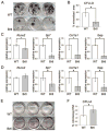
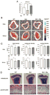
The molecular basis underlying the clinical phenotype in bone diseases is customarily associated with abnormal extracellular matrix structure and/or properties. More recently, cellular malfunction has been identified as a concomitant causative factor and increased attention has focused on stem cells differentiation. Classic osteogenesis imperfecta (OI) is a prototype for heritable bone dysplasias: it has dominant genetic transmission and is caused by mutations in the genes coding for collagen I, the most abundant protein in bone. Using the Brtl mouse, a well-characterized knockin model for moderately severe dominant OI, we demonstrated an impairment in the differentiation of bone marrow progenitor cells toward osteoblasts. In mutant mesenchymal stem cells (MSCs), the expression of early (Runx2 and Sp7) and late (Col1a1 and Ibsp) osteoblastic markers was significantly reduced with respect to wild type (WT). Conversely, mutant MSCs generated more colony-forming unit-adipocytes compared to WT, with more adipocytes per colony, and increased number and size of triglyceride drops per cell. Autophagy upregulation was also demonstrated in mutant adult MSCs differentiating toward osteogenic lineage as consequence of endoplasmic reticulum stress due to mutant collagen retention. Treatment of the Brtl mice with the proteasome inhibitor Bortezomib ameliorated both osteoblast differentiation in vitro and bone properties in vivo as demonstrated by colony-forming unit-osteoblasts assay and peripheral quantitative computed tomography analysis on long bones, respectively. This is the first report of impaired MSC differentiation to osteoblasts in OI, and it identifies a new potential target for the pharmacological treatment of the disorder.


Conflict of interest statement

Disclosure of Potential Conflicts of Interest

The authors indicate no conflict of interest.

Figures

Figure 1
Figure 1
Characterization of bone marrow mesenchymal stem cells (MSCs). (A): Giemsa stained colony-forming unit-fibroblasts (CFU-F). (B): CFU-F quantitation. (C): Examples of fluorescence-activated cell sorting analysis of mesenchymal/progenitor stem cells. Top: Lin, CD31, CD117, CD45, and Ter119 population. Middle: among the previous population CD29+ and CD49e+ cells. Bottom: among the previous population, CD105+ cells in red, control unstained cells in white. (D): Proliferation analysis. ◆ Brtl MSCs, □ WT MSCs. Abbreviation: WT, wild type.
Figure 2
Figure 2
Mesenchymal stem cell (MSC) osteoblastic and adipocytic differentiation. (A): CFU-O Von Kossa staining. (B): CFU-O quantitation. (C, D): Expression level of early (Runx2 and Sp7) and late (Col1a1 and Ibsp) osteoblast-specific genes in (C) MSCs induced toward osteoblastic differentiation and in (D) undifferentiated MSCs. (E): CFU-A oil red O staining. (F): CFU-A quantitation. *, p < .01; #, p < .05. Abbreviations: CFU-A, colony-forming unit-adipogenic; CFU-adipocytes CFU-O, CFU-osteoblasts; WT, wild type.
Figure 3
Figure 3
Mutant collagen retention in the endoplasmic reticulum (ER). (A): Representative Western blot of HSP47. (B): Densitometric analysis of the expression of the ER stress-related marker HSP47 in mesenchymal stem cells (MSCs) differentiated toward the osteoblastic lineage. *, p < .05. (C): Double fluorescence using antibodies against HSP47 (green) and type I collagen (red) in MSCs differentiated toward osteoblast lineage. Green bars represent some of the selected points of colocalization shown below in the graphs. ×40 magnification. Abbreviations: HSP47, heat shock protein 47; WT, wild type.
Figure 4
Figure 4
Expression analysis of autophagy markers. (A): Representative Western blots. (B): Densitometric analysis of the expression levels of autophagic markers in mesenchymal stem cells (MSCs) differentiated toward osteoblasts. (C): Expression analysis of CtsK by qPCR in MSCs differentiated toward osteoblasts. *, p < .05. Abbreviations: ATG7, autophagy-related gene 7; BECN1, Beclin1; CtsK, Cathepsin K; LC3-II, microtubule-associated protein1 light chain 3; qPCR, Quantitative Polymerase Chain Reaction.
Figure 5
Figure 5
Bortezomib effect on cultured bone marrow mesenchymal stem cells (MSCs) and on in vivo bone properties. (A): Quantitation of CFU-O from MSCs of untreated WT (−) and treated (+) and untreated (−) Brtl mice. (B): Representative peripheral quantitative computed tomography metaphyseal scans. The right panel shows the scale of volumetric mineral density. (C): Quantitation of bone density.*, p < .05. (D): Representative hematoxylin and eosin stained sections of tibia metaphysis (top, ×2.5 magnification) and growth plate (bottom, ×25 magnification). Abbreviations: CFU-O, colony-forming unit-osteoblasts; WT, wild type.
Figure 6
Figure 6
Model proposed for MSC impairment in osteogenic differentiation. We propose that preosteoblasts that are not able to complete differentiation, probably due to collagen retention and endoplasmic reticulum stress responsible for autophagy upregulation, either undergo adipogenic lineage or undergo a different unknown fate. Abbreviations: MSC, mesenchymal stem cell; WT, wild type.

References

    1. Forlino A, Cabral WA, Barnes AM, et al. New perspectives on osteogenesis imperfecta. Nat Rev Endocrinol. 2011;7:540–557. - PMC - PubMed
    1. Marini JC, Forlino A, Cabral WA, et al. Consortium for osteogenesis imperfecta mutations in the helical domain of type I collagen: Regions rich in lethal mutations align with collagen binding sites for integrins and proteoglycans. Hum Mutat. 2007;28:209–221. - PMC - PubMed
    1. Forlino A, Porter FD, Lee EJ, et al. Use of the Cre/lox recombination system to develop a non-lethal knock-in murine model for osteogenesis imperfecta with an alpha1(I) G349C substitution. Variability in phenotype in BrtlIV Mice. J Biol Chem. 1999;274:37923–37931. - PubMed
    1. Sillence DO, Senn A, Danks DM. Genetic heterogeneity in osteogenesis imperfecta. J Med Genet. 1979;16:101–116. - PMC - PubMed
    1. Kozloff KM, Carden A, Bergwitz C, et al. Brittle IV mouse model for osteogenesis imperfecta IV demonstrates postpubertal adaptations to improve whole bone strength. J Bone Miner Res. 2004;19:614–622. - PubMed

Publication types

MeSH terms